Search results (98 results)
-
 Bilateral Optic Nerve Pits - ON OCTs
Sep 18 2012 by Pauline T Merrill, MD, FASRS
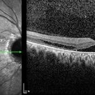
Optic nerve OCTs of a 77-year-old woman with bilateral optic nerve pits and glaucoma, stable over 20 years.
Photographer: Karen Parque, Illinois Retina Associates, Chicago, IL
Imaging device: Zeiss Cirrus
Condition/keywords: optic nerve pit
-
 Binder4 P25 Slide140
Feb 20 2013 by From the Collections of Thomas M. Aaberg, MD and Thomas M. Aaberg Jr., MD
40-year-old white female.
Condition/keywords: optic nerve pit
-
 OCT Image of Optic Nerve Pit and Retinal Detachment
Aug 24 2015 by Young Hee Yoon, MD, PhD
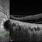
OCT image of a 44-year-old man. There was optic nerve pit and associated foveal detachment. His best-corrected visual acuity was count finger in 30cm.
Photographer: Jung Im Cho, Asan Medical Center
Imaging device: Spectralis OCT
Condition/keywords: optic nerve pit
-
 OCT Image of Optic Nerve Pit and Retinal Detachment
Aug 24 2015 by Young Hee Yoon, MD, PhD
SD-OCT image of a 44-year-old man. There was optic nerve pit and associated foveal detachment. His best-corrected visual acuity was count finger in 30cm.
Photographer: Jung Im Cho, Asan Medical Center
Imaging device: Spectralis OCT
Condition/keywords: optic nerve pit
-
 OCT Image of Optic Nerve Pit and Retinal Detachment
Aug 24 2015 by Young Hee Yoon, MD, PhD
OCT image of a 44-year-old man. There was optic nerve pit and associated foveal detachment. His best-corrected visual acuity was count finger in 30cm.
Photographer: Jung Im Cho, Asan Medical Center
Imaging device: Spectralis OCT
Condition/keywords: optic nerve pit
-
 Optic Disc Pit
Nov 8 2021 by Michael Grinton
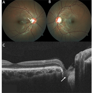
Optic disc pits are rare congenital abnormalities of the optic nerve head. Colour fundus image of an asymptomatic 18-year old male shows an optic disc pit in the right eye (A, white arrow); a small, grey, oval shaped excavation in the temporal segment of the optic disc. These pits are usually unilateral (B shows normal colour fundus of left eye) and asymptomatic. Imaging with optical coherence tomography (C) shows the optic disc pit in cross section (white arrow) and normal macular structure. In some patients with the condition, fluid can accumulate underneath the macular (serous macular detachment).
Condition/keywords: Optic disc pit, Optic nerve pit, Optic pit
-
 Optic Nerve Head Pit
Feb 20 2013 by From the Collections of Thomas M. Aaberg, MD and Thomas M. Aaberg Jr., MD
Optic nerve head pit Red free photograph
Condition/keywords: optic nerve pit
-
 Optic Nerve Head Pit
Feb 20 2013 by From the Collections of Thomas M. Aaberg, MD and Thomas M. Aaberg Jr., MD
optic nerve head pit Red free photograph
Condition/keywords: optic nerve pit
-
 Optic Nerve Head Pit with Fluid
Feb 20 2013 by From the Collections of Thomas M. Aaberg, MD and Thomas M. Aaberg Jr., MD
Optic Nerve Head Pit Red Free Photo
Condition/keywords: optic nerve pit, red-free
-
 Optic Nerve Pit
Feb 21 2024 by Virginia Gebhart
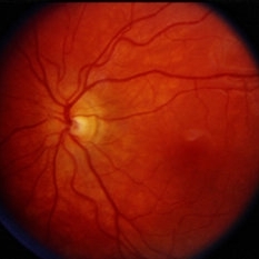
65 year old female with optic nerve pit. Asymptomatic, continued observation.
Photographer: Virginia Gebhart
Imaging device: Topcon TRC 50DX
Condition/keywords: congenital optic nerve pit, Optic nerve pit
-
 Optic Nerve Pit
Feb 20 2013 by From the Collections of Thomas M. Aaberg, MD and Thomas M. Aaberg Jr., MD
No history.
Condition/keywords: optic nerve pit
-
 Optic Nerve Pit
Feb 20 2013 by From the Collections of Thomas M. Aaberg, MD and Thomas M. Aaberg Jr., MD
No history; FA.
Condition/keywords: optic nerve pit
-
 Optic Nerve Pit
Feb 20 2013 by From the Collections of Thomas M. Aaberg, MD and Thomas M. Aaberg Jr., MD
No history; FA; no leak.
Condition/keywords: optic nerve pit
-
 Optic Nerve Pit
Feb 19 2013 by From the Collections of Thomas M. Aaberg, MD and Thomas M. Aaberg Jr., MD
Optic nerve pit with serous macular detachment.
Condition/keywords: macular detachment, optic nerve pit
-
 Optic Nerve Pit
Dec 11 2014 by H. Michael Lambert, MD
High mag LE color photo of ON pit.
Condition/keywords: optic nerve pit
-
 Optic Nerve Pit
Dec 11 2014 by H. Michael Lambert, MD
High mag RE color photo of ON pit.
Condition/keywords: optic nerve pit
-
 Optic Nerve Pit
Dec 11 2014 by H. Michael Lambert, MD
Fluorescein angiogram of ON pit LE.
Condition/keywords: optic nerve pit
-
 Optic Nerve Pit
Aug 30 2012 by Raj K. Maturi, MD
Photographer: Tom Steele, CRA, Midwest Eye Institute
Imaging device: Topcon Ex
Condition/keywords: optic nerve pit
-
 Optic Nerve Pit
Aug 30 2012 by Raj K. Maturi, MD
congenital optic nerve pit with chronic pigment changes in macula due to detachment
Photographer: Tom Steele, CRA, Midwest Eye Institute
Imaging device: Topcon Ex
Condition/keywords: optic nerve pit
-
 Optic Nerve Pit
Aug 30 2012 by Raj K. Maturi, MD
Photographer: Tom Steele, CRA, Midwest Eye Institute
Imaging device: Topcon Ex
Condition/keywords: optic nerve pit
-
 Optic Nerve Pit
Aug 30 2012 by Raj K. Maturi, MD
Photographer: Tom Steele, CRA, Midwest Eye Institute
Imaging device: Topcon Ex
Condition/keywords: optic nerve pit
-
 Optic Nerve Pit
Aug 30 2012 by Raj K. Maturi, MD
Photographer: Tom Steele, CRA, Midwest Eye Institute
Imaging device: Topcon Ex
Condition/keywords: optic nerve pit
-
 Optic Nerve Pit
Aug 2 2018 by Nilesh K Kanjani, MD
Fundus photograph of 45-year-old woman with complain of gradual progressive dimness of vision in LE.
Photographer: Dr Nilesh Kanjani
Condition/keywords: optic nerve pit
-
 Optic Nerve Pit
Aug 4 2018 by Nilesh K Kanjani, MD
A female patient aged 40 years came with progressive dimness of vision.
Photographer: Dr Nilesh Kanjani
Condition/keywords: optic nerve pit
-
 Optic Nerve Pit
Feb 19 2013 by From the Collections of Thomas M. Aaberg, MD and Thomas M. Aaberg Jr., MD
Left side of a stereo pair.
Condition/keywords: avulsed epiretinal membrane, optic nerve pit, stereo pair
 Loading…