Search results (9 results)
-
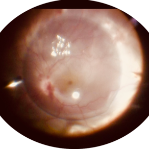
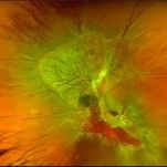 Diabetic TRD - Photos OS
Nov 17 2019 by John S. King, MD
28-year-old white male with poorly controlled Type 1 DM, with a history of non-compliance with follow-ups, was referred with for DR with CME OS, and 3 weeks decrease vision OS. Va cc was 20/15 OD and HM OS. IOP 18/14. No NVI OU. Posteriorly, the right eye had macular exudates, no NVD, and a large area of NVE along the IT arcade. The left eye large NV plaque around disc, wrapping macula, with total RD with a posterior funnel appearance. The FA in the left eye showed severe peripheral and macular ischemia with diffuse leakage from a fibrovascualr plaque.
Photographer: Adriana Shelby
Imaging device: Optos CA
Condition/keywords: diabetic traction detachment, open funnel RD
-
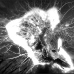 Diabetic TRD OS - FA 1 Min
Nov 17 2019 by John S. King, MD
28-year-old white male with poorly controlled Type 1 DM, with a history of non-compliance with follow-ups, was referred with for DR with CME OS, and 3 weeks decrease vision OS. Va cc was 20/15 OD and HM OS. IOP 18/14. No NVI OU. Posteriorly, the right eye had macular exudates, no NVD, and a large area of NVE along the IT arcade. The left eye large NV plaque around disc, wrapping macula, with total RD with a posterior funnel appearance. The FA in the left eye showed severe peripheral and macular ischemia with diffuse leakage from a fibrovascualr plaque.
Photographer: Adriana Shelby
Imaging device: Optos CA
Condition/keywords: diabetic traction detachment, open funnel RD
-
 Eye Finally Got the Ring... But the Retina Was Too Detached to Care
Nov 5 2025 by SHRADDHA RAJ SHRIVASTAVA
Left Eye B-scan ultrasound of a patient with old retinal detachment shows open funnel shaped hyperechoic membranous echoes, with high amplitude spikes on A-scan and a poor after-movement on dynamic B-scan, suggestive of retinal detachment. We can see a round echogenicity in sub-retinal location, with clear contents within, suggestive of a retinal cyst. This B-scan image is indicative of a long-standing chronic retinal detachment with secondary retinal cyst.
Photographer: Dr. Shraddha Raj Shrivastava
Condition/keywords: B scan ultrasound, chronic retinal detachment, OLD RD, open funnel RD, retinal cyst
-
 Funnel Retinal Detachment
Jun 11 2023 by Ethan K Sobol, MD
Intraoperative view of a funnel retinal detachment with proliferative vitreoretinoapthy in an eye with previous open globe injury. PVR membranes were peeled, and the retina was flattened and re-attached with an inferior relaxing retinotomy and silicone oil tamponade
Condition/keywords: intraoperative, open funnel RD, open globe injury, proliferative vitreoretinopathy (PVR)
-
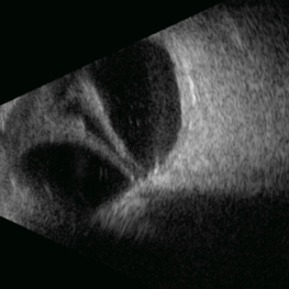 Open Funnel
Apr 10 2025 by Gustavo Uriel Fonseca Aguirre
This B-mode longitudinal ultrasound scan demonstrates a long-standing rhegmatogenous retinal detachment, showing a characteristic open funnel configuration. The findings are consistent with chronic retinal detachment.
Photographer: Gustavo U. Fonseca Aguirre, Hospital Conde de Valenciana, Ciudad de México
Imaging device: Funnel
Condition/keywords: open funnel RD, Retina detachment
-
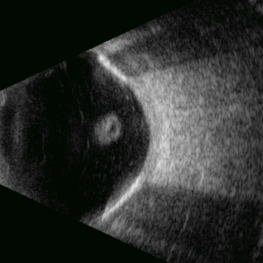 Open Funnel (Transversal)
Apr 10 2025 by Gustavo Uriel Fonseca Aguirre
This B-mode transverse ultrasound scan reveals a chronic rhegmatogenous retinal detachment, demonstrating a funnel-shaped configuration with a narrow intraluminal space. Two hyperechoic choroidal calcifications are present, indicative of chronicity.
Photographer: Gustavo U. Fonseca Aguirre, Hospital Conde de Valenciana, Ciudad de México
Condition/keywords: open funnel RD, Retina detachment
-
 Open Funnel Retinal Detachment
Oct 13 2012 by Geoffrey G. Emerson, MD, PhD, FASRS
Open funnel retinal detachment
Condition/keywords: B scan ultrasound, open funnel RD
-
 Retinal Detachment
Oct 29 2023 by Anand Temkar
Membranous echoes with high spikes with restricted after movements suggestive of retinal detachmment.
Photographer: Dr.Anand Temkar- Retina Foundation, Ahmedabad
Condition/keywords: A-scan ultrasound, B scan ultrasound, open funnel RD
-
 PFC- Thor's Mjolnir in RD cases.
Jul 25 2020 by SANDEEP KUMAR
PFC in chronic retinal detachment has a vital role. The image is of a 12-year-old girl with open funnel RD. Characteristically PFCLs have high specific gravity ranging from 1.76 to 2.03, low surface tension, and viscosity. These physical properties make perfluorocarbon liquids an ideal for intraoperative tool in vitreoretinal surgery. With high specific gravity they flatten the detcahed retina and push the SRF anteriorly giving surgeon room for maneuvers.
Photographer: Dr Daraius Shroff . Shroff Eye Centre New Delhi
Condition/keywords: perfluorooctane
 Loading…