Search results (30 results)
-
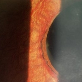
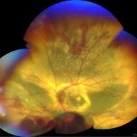
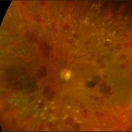
 Advanced Coats' Disease with Neovascular Glaucoma
Aug 21 2019 by Victor M Villegas, MD
Advanced Coats' Disease with neovascular glaucoma.
Photographer: Giselle Deoliveira, Bascom Palmer Eye Institute, University of Miami
Imaging device: RetCam III
Condition/keywords: abnormal retinal vessel, bullous retinal detachment, Coats' disease, diffuse lipid exudate, foveal hard exudates, neovascular glaucoma, pediatric retina
-
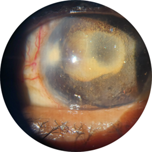
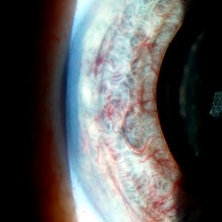
 Advanced Stage of Neovascular Glaucoma
Mar 21 2013 by Yusuke Oshima, MD, PhD
An 82-year-old man with a advanced stage of neovascular glaucoma. A slit-lamp photograph illustrates iris ectropion with prominent iris neovascularization.
Photographer: Yusuke Takada, Osaka University Graduate School of Medicine
Condition/keywords: neovascular glaucoma
-
 Angle Closure Glaucoma
Nov 14 2019 by Jennifer Schiefer, CRA, COA
96-year-old female who presented in clinic with eye pain x 3 weeks and HM Va. Unique case in pseudophakic eye. Exam showed sectoral area of NVI. Questionable NVG after RVO.
Photographer: Jennifer Schiefer
Condition/keywords: angle closure, anterior chamber, neovascular glaucoma
-
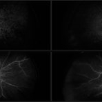
 Central Retinal Vein Occlusion with Retinal Neovascularization
Jan 19 2022 by Olivia Rainey
Ultra-widefield fluorescein angiogram of a 56-year-old male with a Central Retinal Vein Occlusion with Retinal Neovascularization affecting his left eye. The patient presented on 1/19/2022 with scNLP vision in the left eye. The patient has good PRP, however areas of ischemia still remain untreated by laser. He also has severe neovascular glaucoma contributing to his poor vision.
Photographer: Olivia Rainey, OCT-C, COA
Imaging device: Optos California
Condition/keywords: central retinal vein occlusion (CRVO), FA early phase, fluorescein angiogram (FA), hemorrhage, ischemic CRVO, left eye, neovascular glaucoma, Optos, pan-retinal photocoagulation (PRP), retinal ischemia, retinal neovascularization, ultra-wide field imaging
-
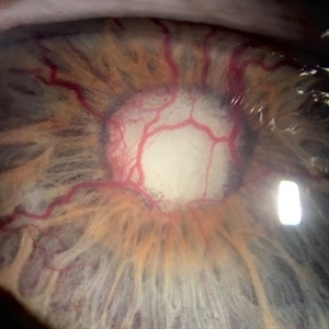
 Neovascular Glaucoma
Jan 3 2020 by Manuel Ángel Alcántara Delgado, MD
Slit lamp photograph of a 65-year-old man with diabetic retinopathy and previous history of phaco-vitrectomy.
Photographer: Manuel Ángel Alcántara Delgado, CMN SXXI, Mexico City
Condition/keywords: diabetes, diabetic retinopathy, neovascular glaucoma, neovascularization (NV), retina surgery, retina surgery complications
-
 Neovascular Glaucoma
Jan 4 2020 by Manuel Ángel Alcántara Delgado, MD
Slit lamp photograph of a 69-year-old man with diabetic retinopathy and poor metabolic control.
Photographer: Manuel Ángel Alcántara Delgado, CMN SXXI, Mexico City.
Condition/keywords: diabetes, diabetic retinopathy vitrectomy study (DRVS), neovascular glaucoma
-
 Neovascular Glaucoma
Dec 4 2015 by Kathy Karsten, COT
Ahmad tube shunt with peaked pupil in the left eye of a 63-year-old woman for neovascular glaucoma.
Photographer: Kathy Karsten, COT
Imaging device: Topcon TRC 50-DX
Condition/keywords: angle neovascularization
-
 Neovascular Glaucoma Tree
Jun 23 2021 by Ana Karen Lopez, MD
Anterior segment photography of an 54-year-old man with neovascular glaucoma.
Photographer: Ana Karen López-Vázquez, MD
Condition/keywords: angle neovascularization, anterior chamber, cataract with neovascularization, neovascular glaucoma, neovascularization (NV), neovascularization of iris (NVI)
-
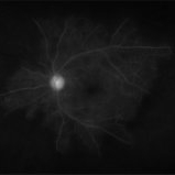
 Neovascularization Glaucoma
Jul 14 2013 by Jason S. Calhoun
Fundus photo shows abnormal blood vessels growing in the optic disc.
Photographer: Jason S. Calhoun, Department of Ophthalmology, Mayo Clinic Jacksonville, Florida
Imaging device: TOPCON TRC 50-EX
Condition/keywords: neovascular glaucoma
-
 NVI in a 50-Year-Old with NVG and Peripheral Retinal Occlusion
Apr 21 2020 by Theodore Leng, MD, MS, FASRS
NVI in a 50-year-old with NVG and peripheral retinal occlusion
Condition/keywords: neovascular glaucoma, neovascularization of iris (NVI)
-
 Optic Cupping With Neovascularization
Jul 14 2013 by Jason S. Calhoun
Fundus photo shows abnormal blood vessels growing in the optic disc with severe glaucomatous cupping.
Photographer: Jason S. Calhoun, Department of Ophthalmology, Mayo Clinic Jacksonville, Florida
Imaging device: TOPCON TRC 50-EX
Condition/keywords: neovascular glaucoma
-
 Proliferative Diabetic Retinopathy with Neovascular Glaucoma
Jul 12 2018 by Nichole Lewis
68-year-old male with proliferative diabetic retinopathy, severe capillary nonperfusion and neovascular glaucoma. Treated with Avastin intra-ocular injection and future pan-retinal photocoagulation. VA 20/300.
Photographer: Nichole Lewis
Condition/keywords: capillary nonperfusion, diabetes, neovascular glaucoma, non-perfusion, proliferative diabetic retinopathy (PDR)
-
 Rubeosis Iridis
Jul 21 2025 by Luai Abu-Ismail, MD
73y old female patient, known case of uncontrolled DM for more than 15y and chronic kidney disease. She had CRVO and complicated with 100 day neovascularization glaucoma.
Photographer: Luai Abu-Ismail
Imaging device: S23 Ultra
Condition/keywords: central retinal vein occlusion (CRVO), neovascular glaucoma, Rubeosis
-
 Severe Rubeosis and Angle Neovascularization
Nov 21 2019 by Anfisa Ayalon, MD
Patient with proliferative diabetic retinopathy, severe retinal ischemia, rubeosis and angle neovascularization with IOP elevation.
Photographer: Anfisa Ayalon,MD., Meir Medical Center, Kfar Saba, Israel.
Imaging device: with gonioscopy lens
Condition/keywords: angle neovascularization, neovascular glaucoma, proliferative diabetic retinopathy (PDR), rubeosis
-
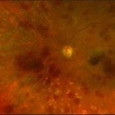
 Coats' disease
Sep 7 2022 by Niloofar Piri, MD
Total exudative RD with extensive subretinal exudates and peripheral telangiectatic vascular anomalies in stage 4 Coats's disease. Patient is a 12 yo who presented with severe eye pain and neovascular glaucoma secondary to the above.
Photographer: Jacob Grodsky, MD
Condition/keywords: Coats' disease
-
 Crystallized silicone oil particles in the anterior chamber
Oct 26 2023 by Anmol Naik, MS, DNB, FMRF, FICO, MNAMS
Anterior segment image of a 67-year-old Indian woman who had proliferative diabetic retinopathy with traction retinal detachment with neovascular glaucoma. Patient underwent vitrectomy with membrane peeling with endolaser followed by silicone oil injection 1 year back. Patient was lost to follow up and presented a year later with this picture. She had crystallized silicone oil particles in the anterior chamber rendering a polychromatic lustre like appearance; a unique and rare finding.
Photographer: Anmol Naik
Condition/keywords: Polychromatic lustre in Anterior Chamber
-
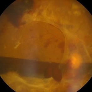
 Endoscopy: Peripheral Endoscopic Ciliary Body Ablation for Control of Neovascular Glaucoma in Diabetic Patient
Dec 10 2012 by Yale L. Fisher, MD
With this patient microscopic control was not possible due to a small fixed pupil with vascularized synechiae to a posterior chamber IOL. There was recurrent intraocular bleeding and elevated IOP. The ciliary body ablation was accomplished first with a 532 laser fed through the working channel of the endoscope. Examination of the peripheral retina revealed a ring-like rhegmatogenous retinal separation and a large inferior tear with persistent traction. Endoscopically controlled imaging and a second instrument (suction/cutter) removed the tractional elements and permitted an air fluid exchange. Retinal reattachment occurred as the air-fluid exchange was completed permitting laser ablation of the ring like area that had been separated. The entire procedure was performed utilizing the small gauge endoscope.
Condition/keywords: video
-
 Monocular, Proliferative Diabetic Retinopathy
Sep 8 2021 by VERONICA ADRIANA ROMERO- MORALES, MD
Fundus photograph of a 70-year-old man with proliferative diabetic retinopathy and neovascular glaucoma. BCVA 20/200 right eye.
Photographer: Lic. Belgica Copado Andrade
Imaging device: Cobra HD
Condition/keywords: fibrovascular proliferation, ischaemic diabetic maculopathy, proliferative diabetic retinopathy (PDR), tractional retinal detachment
-
 Ocular Ischemia From Left Carotid Artery Occlusion
Jan 8 2015 by Connie J Chen, MD
Fundus photo of a 66-year-old female with insulin dependent diabetes. She presented with eye pain due to neovascular glaucoma and angiography demonstrates extensive non-perfusion in the left eye with leakage from neovascularization of the disc. The right eye is completely perfused with no neovascular changes
Photographer: David Emmert
Imaging device: Optos Widefield Angiography
Condition/keywords: non-perfusion, ocular ischemic syndrome
-
 Ocular Ischemic Symdrome
Jan 27 2018 by Alex H. Rubowitz, MD
A 65-year-old male with known diabetic retinopathy presented with iris rubeosis and neovascular glaucoma in his left eye. Photo was taken prior to his secind laser PRP session in the eye, and shows typical round hemorrhages of OIS. A systemic workup revealed severe carotid stenosis.
Photographer: Lilach, Meir Hospital Retina Clinic
Imaging device: Optos California
Condition/keywords: ocular ischemic syndrome
-
 Ocular Ischemic Syndrome
Jan 27 2018 by Alex H. Rubowitz, MD
A 65-year-old male with known diabetic retinopathy presented with iris rubeosis and neovascular glaucoma in his left eye. Photo was taken prior to his secind laser PRP session in the eye, and shows typical round hemorrhages of OIS. A systemic workup revealed severe carotid stenosis.
Photographer: Lilach, Meir Hospital Retina Clinic
Imaging device: Optos California
Condition/keywords: ocular ischemic syndrome
-
 Ocular Ischemic Syndrome With Neovascularization Due to Left Cartoid Artery Occlusion
Jan 8 2015 by Connie J Chen, MD
Fundus photo of a 66-year-old female with insulin dependent diabetes. She presented with eye pain due to neovascular glaucoma and angiography demonstrates extensive non-perfusion in the left eye with leakage from neovascularization of the disc. The right eye is completely perfused with no neovascular changes
Photographer: David Emmert
Imaging device: Optos Widefield Angiography
Condition/keywords: non-perfusion, ocular ischemic syndrome
-
 Retinal Ischemia Secondary to Diabetic Retinopathy
Aug 29 2018 by Olivia Rainey
Fluorescein angiogram series of a 57-year-old male patient with proliferative diabetic retinopathy of the right eye. Patient has delayed AV transit with significant retinal ischemia and retinal capillary nonperfusion. The ischemia is extensive resulting in neovascularization of the iris and consequently neovascular glaucoma.
Photographer: Olivia Rainey
Imaging device: Optos
Condition/keywords: diabetes, disc hyperfluorescene, fluorescein angiogram (FA), non-perfusion, Optos, proliferative diabetic retinopathy (PDR), retinal ischemia, ultra-wide field imaging, vitreous hemorrhage
-
 Rubeosis Iridis
Jul 8 2013 by Jason S. Calhoun
Patient with rubeosis iridis in the right eye due to neovascular glaucoma. VA is 20/40 in the right eye. Will follow up in 3 months.
Photographer: Jason S. Calhoun, Department of Ophthalmology, Mayo Clinic Jacksonville, Florida
-
---thumb.JPG/image-square;max$300,300.ImageHandler) Rubeosis Iridis
Rubeosis Iridis
Jul 8 2013 by Jason S. Calhoun
Patient with rubeosis iridis in the right eye due to neovascular glaucoma. VA is 20/40 in the right eye. Will follow up in 3 months.
Photographer: Jason S. Calhoun, Department of Ophthalmology, Mayo Clinic Jacksonville, Florida
 Loading…