Search results (26 results)
-
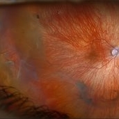
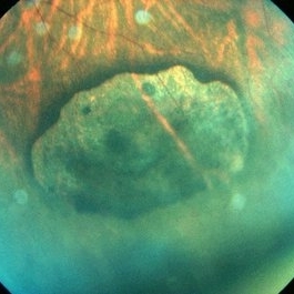 CHRPE
CHRPE
May 9 2014 by S. Natarajan, MD, FASRS, FRCS (GLASGOW) , FICO, D.Sc, FELA
23-year-old male in for routine eye checkup with BCVA 6/6 (OU). Showed CHRPE lesion in INQ (OD )on fundus.
Photographer: ADITYA JYOT EYE HOSPITAL,MUMBAI,INDIA
Condition/keywords: congenital hypertrophy of the retinal pigment epithelium (CHRPE), myopic eye
-
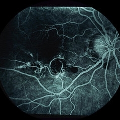
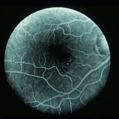
 CNVM - Myopic
-
 CNVM - Myopic
-
 CNVM - Myopic
-
 CNVM - Myopic
-
 CNVM - Myopic
-
 CNVM - Myopic
-
 CNVM - Myopic
-
 CNVM - Myopic
-
 CNVM - Myopic
-
 CNVM - Myopic
-
 CNVM - Myopic
-
 CNVM - Myopic
-
 CNVM - Myopic
-
 CNVM - Myopic
-
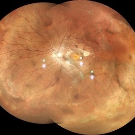
 Macular Hole Retinal Detachment Over a Posterior Staphyloma
Dec 31 2016 by Linda A Cernichiaro- Espinosa, MD
Macular hole retinal detachment over a posterior staphyloma of pathologic myopia.
Photographer: Linda A Cernichiaro
Imaging device: Optos
Condition/keywords: degenerative myopia, high myopia, macular hole, myopic eye, posterior staphyloma, vitreoretinal degeneration
-
 Pathological Myopia
Oct 18 2023 by Anand Temkar
LE widefield CF montage of a 24 year old male with pathological myopia showing multiple lattice degenerations in periphery along with holes.
Photographer: Dr.Anand Temkar- Retina Foundation, Ahmedabad
Imaging device: Mirante
Condition/keywords: holes, lattice degeneration, myopic degeneration, myopic eye
-
 Pseudo Retinal Break
Nov 9 2012 by Norman Byer
This 23-year-old man presented with a fresh retinal detachment in a highly myopic eye and this very unusual retinal appearance. You can see two reddish areas with fairly distinct borders which at first make us think of retinal breaks. However, the left area has two tiny vessels visible in it, and the right area shows visible translucent retinal tissue extending across it. This patient has extensive areas of paving stone degeneration. Usually, such lesions present a barrier to a detaching retina and areas of paving stone usually remain attached. However, in this photograph we can see two paving stone lesions, and the detachment has extended right through them peeling them off from the underlying pigment epithelium. The two reddish areas, therefore, represent the very thin retina which previously constituted part of two paving stone lesions. The yellow atrophic areas which are visible deep to the detached retina represent the deeper parts of the same two original paving stone lesions.
Condition/keywords: myopic eye, pigment epithelium, reddish lesion, yellow atrophic area
-
 Retinal Detachment
Mar 5 2025 by Kimberly Wakester
Optomap RGB image of an 9-year-old boy with a retinal detachment with retinal break at 9:00 in the right eye. Surgery was recommended. Patient is to continue follow up care post operatively.
Photographer: Kimberly Wakester, COA
Imaging device: Optos California
Condition/keywords: myopic eye, Retinal Detachment, retinal tear
-
 IOL With BAG in Vitreous of Myopic Eye
Apr 14 2017 by Manish Nagpal, MD, FRCS (UK), FASRS
50-year-old male having myopia presented with a IOL in vitreous within its bag.
Photographer: Pooja Barot
Condition/keywords: intraocular lens (IOL), intraocular lense in vitreous, intraocular lense with bag, myopia
-
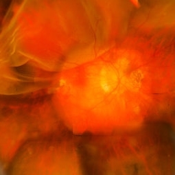
 Pan-retinal Photocoagulation in a Myopic Eye
Aug 27 2013 by Carmen L Gonzalez, MD
Ultra-wide-field fundus photograph of a myopic patient.
Photographer: Regina Victoria, Denver Health Medical Center, Denver, Colorado
Imaging device: Optomap, Panoramic 200; Optos PLC, Scotland , UK
Condition/keywords: pan-retinal photocoagulation (PRP)
-
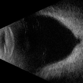 Posterior Staphyloma + ON-Coloboma
Aug 20 2025 by Gustavo Uriel Fonseca Aguirre
This axial B-scan reveals a highly myopic eye with a posterior staphyloma and an associated optic nerve coloboma. The staphyloma appears as a deep scleral outpouching adjacent to the optic disc, while the coloboma demonstrates a focal posterior excavation with retrobulbar extension.
Photographer: Gustavo U. Fonseca Aguirre, Hospital Conde de Valenciana, Ciudad de México
Condition/keywords: optic nerve coloboma, posterior staphyloma
-
 Retinal Detachment
Feb 24 2017 by Manish Nagpal, MD, FRCS (UK), FASRS
Patient presenting with a fresh superior retinal detachment in a myopic eye.
Photographer: Pooja Barot
-
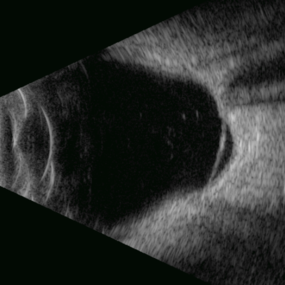 Retinal Detachment Associated With a Posterior Staphyloma
Apr 9 2025 by Gustavo Uriel Fonseca Aguirre
B-mode axial ultrasound scan of a highly myopic eye shows a posterior staphyloma with an associated macular hole-induced retinal detachment.
Photographer: Gustavo U. Fonseca Aguirre, Hospital Conde de Valenciana, Ciudad de México
Condition/keywords: high myopia, posterior staphyloma, rhegmatogenous retinal detachment
-
 RRD in Posterior Staphyloma
May 21 2025 by Gustavo Uriel Fonseca Aguirre
This B-mode axial ultrasound scan of a highly myopic eye demonstrates a prominent posterior staphyloma with an associated inferior retinal detachment sparing the macular region.
Photographer: Gustavo U. Fonseca Aguirre, Hospital Conde de Valenciana, Ciudad de México
Condition/keywords: high myopia, posterior staphyloma, Retina detachment
 Loading…