Search results (80 results)
-
 Coats' Disease
Mar 4 2017 by Hashim Ali Khan, OD, FAAO
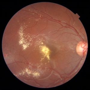
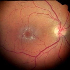
Color Fundus Image of an 18-year-old girl with Coats disease.
Condition/keywords: Coats' disease, exudates over the posterior pole, macular edema, macular telangiectasia
-
 IMT2 With Scaring
Sep 19 2017 by Theodore Leng, MD, MS, FASRS
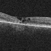
IMT2 with scarring and ILM draping.
Condition/keywords: draping, IMT2, internal limiting membrane (ILM) peeling, macular telangiectasia
-
 Macular Telangiectasia (Auto Fluorescence)
May 16 2014 by Avris Romario Diparaja Siahaan
AF Image of a 58-year-old-man with a macular telangiectasia condition on his left eye.
Photographer: Avris Romario Diparaja Siahaan, Klinik Mata Nusantara
Imaging device: Topcon TRC 50 DX Type IA
Condition/keywords: fundus autofluorescence (FAF), macular telangiectasia
-
 Macular Telangiectasia (FA Early Phase)
May 16 2014 by Avris Romario Diparaja Siahaan
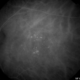
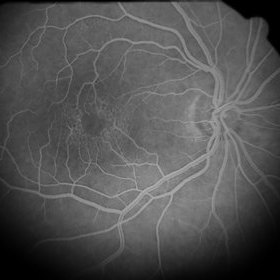
FA (early phase) image of a 58-year-old-man with a macular telangiectasia condition on his left eye.
Photographer: Avris Romario Diparaja Siahaan, Klinik Mata Nusantara
Imaging device: Topcon TRC 50 DX Type IA
Condition/keywords: FA early phase, macular telangiectasia
-
 Macular Telangiectasia (FA Late Phase)
May 16 2014 by Avris Romario Diparaja Siahaan
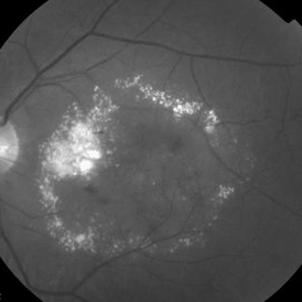
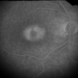
FA (late phase) image of a 58-year-old-man with a macular telangiectasia condition on his left eye.
Photographer: Avris Romario Diparaja Siahaan, Klinik Mata Nusantara
Imaging device: Topcon TRC 50 DX Type IA
Condition/keywords: FA late phase, macular telangiectasia
-
 Macular Telangiectasia (ICG Early Phase)
May 16 2014 by Avris Romario Diparaja Siahaan
ICGA (early phase) image of a 58-year-old-man with a macular telangiectasia condition on his left eye.
Photographer: Avris Romario Diparaja Siahaan, Klinik Mata Nusantara
Imaging device: Topcon TRC 50 DX Type IA
Condition/keywords: indocyanine green (ICG) angiography, macular telangiectasia
-
 Macular Telangiectasia (ICG Late Phase)
May 16 2014 by Avris Romario Diparaja Siahaan
ICGA image of a 58-year-old-man with a macular telangiectasia condition on his left eye.
Photographer: Avris Romario Diparaja Siahaan, Klinik Mata Nusantara
Imaging device: Topcon TRC 50 DX Type IA
Condition/keywords: indocyanine green (ICG) angiography, macular telangiectasia
-
 Macular Telangiectasia (Red Free)
May 16 2014 by Avris Romario Diparaja Siahaan
A red free image of a 58-year-old-man with a macular telangiectasia condition on his left eye.
Photographer: Avris Romario Diparaja Siahaan, Klinik Mata Nusantara
Imaging device: Topcon TRC 50 DX Type IA
Condition/keywords: macular telangiectasia, red-free
-
 Macular Telangiectasia Type 2
Mar 8 2018 by Daniel R Agarwal, MD
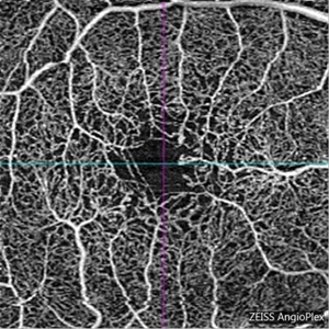
OCT Angiography image in a 51-year-old male with fogging of vision and leaking on fluorescein angiography.
Photographer: Jen Welsh
Imaging device: Zeiss Angioplex OCTA
Condition/keywords: macular telangiectasia, macular telangiectasia type 2
-
 Macular Telangiectasis
May 13 2019 by Hashim Ali Khan, OD, FAAO
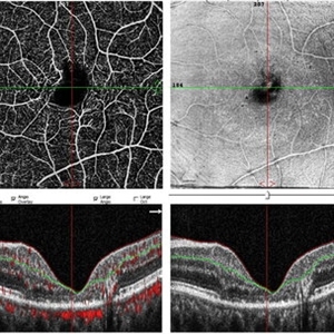
OCT-angio of superficial vascular network and structural OCT of a 60-years-old female demonstrating macular TEL showing alterations in FAZ and vascular remodeling and increased the intercapillary distance.
Imaging device: Optical Coherence Tomography Angiography
Condition/keywords: idiopathic macular telangiectasia, macular telangiectasia, macular telangiectasia type 2
-
 Macular Teleangiectasia
May 29 2013 by Zofia Anna Nawrocka (vel Michalewska), MD, PhD
Late phase fluorescein angiography of a 56-year-old woman.
Photographer: Janusz Michalewski, MD, PhD, Ophthalmic Clinic "Jasne Blonia", Lodz, Poland
Imaging device: Heidelberg Spectralis
Condition/keywords: macular telangiectasia
-
 Macular Teleangiectasia
May 29 2013 by Zofia Anna Nawrocka (vel Michalewska), MD, PhD
Scanning laser ophthalmoscopy and SD-OCT of a 56-year old-woman. Visual acuity was 0.8 Snellen.
Photographer: Janusz Michalewski, MD, PhD, Ophthalmic Clinic "Jasne Blonia", Lodz, Poaldn
Imaging device: Heidelberg Spectralis
Condition/keywords: macular telangiectasia, optical coherence tomography (OCT)
-
 Retinal Crystals in Macular Telangiectasia
Jan 6 2019 by John S. King, MD
61-year-old white female, healthy, slow progressive central vision loss OD. 20/200 OD and 20/30 OS with mild-mod NSC OU. Retinal crystals around fovea in the posterior pole. FA shows 360 degrees of parafoveal telangiectatic vessels that leak without signs of CNVM. Minimal changes in fellow eye. Asymmetric MacTel 2. Monitoring and using AREDS 2.
Photographer: Kay Dalby
Imaging device: Topcon 50 (satellite office)
Condition/keywords: crystalline retinopathy, cystoid macular edema (CME), macular telangiectasia
-
 Type 1A Macular Telangiectasia
Nov 11 2013 by Gerardo Garcia-Aguirre, MD
Type 1A macular telangiectasia.
Condition/keywords: macular telangiectasia
-
 Type 1A Macular Telangiectasia - Autofluorescence
Nov 11 2013 by Gerardo Garcia-Aguirre, MD
Autofluorescence image showing hypoautofluorescent spots corresponding to telangiectatic vessels.
Condition/keywords: macular telangiectasia
-
 Type 1A Macular Telangiectasia - Fluorescein Angiogram - Early
Nov 11 2013 by Gerardo Garcia-Aguirre, MD
Fluorescein angiogram showing hyperfluorescent spots temporal and inferior to the fovea, with mild leakage.
Condition/keywords: macular telangiectasia
-
 Type 1A Macular Telangiectasia - Fluorescein Angiogram - Late
Nov 11 2013 by Gerardo Garcia-Aguirre, MD
Fluorescein angiogram showing hyperfluorescent spots with diffuse leakage.
Condition/keywords: macular telangiectasia
-
 Type 1A Macular Telangiectasia - Fundus photograph
Nov 11 2013 by Gerardo Garcia-Aguirre, MD
Fundus photograph of a 43-year-old male complaining of mild metamorphopsia in OS. BCVA 20/25. Some hard exudates and telangiectatic vessels are observed inferior and temporal to the fovea.
Condition/keywords: macular telangiectasia
-
 Type 1A Macular Telangiectasia - ICG Angiogram
Nov 11 2013 by Gerardo Garcia-Aguirre, MD
ICG Angiogram showing hyperfluorescent spots temporal and inferior to the fovea.
Condition/keywords: indocyanine green (ICG) angiography, macular telangiectasia
-
 Type 1A Macular Telangiectasia - OCT
Nov 11 2013 by Gerardo Garcia-Aguirre, MD
SD-OCT showing intraretinal fluid in both internal and external layers of the retina. Hyper-reflective foci are also visible in the external layers of the retina.
Condition/keywords: macular telangiectasia, optical coherence tomography (OCT)
-
 MACTEL
Mar 7 2025 by T. P . VIGNESH, MBBS,MS
Fundus photograph of the left eye of an 62-year-old woman with macular telangiectasia type 2.
Photographer: Sivanath
Imaging device: EIDON
Condition/keywords: macular telangiectasia type 2
-
 Mactel Type 2
Jul 1 2022 by T. P . VIGNESH, MBBS,MS
Fundus photo of a 70 year old man with Mactel with scarred subretinal neovascular membrane.
Photographer: Bharathi Singaravel
Imaging device: Zeiss Clarus
Condition/keywords: macular telangiectasia type 2
 Loading…