Search results (9 results)
-
 Intraocular lens luxated to the vitreous cavity
Jun 24 2023 by Mariam Cernichiaro-Espinosa, MD
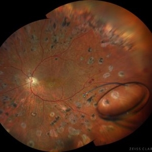
Three-piece intraocular lens luxated to the vitreous cavity in a patient with photocoagulated diabetic retinopathy after blunt trauma.
Photographer: Mariam Cernichiaro-Espinosa, Asociación para Evitar la Ceguera en México, I.A.P. Mexico City, Mexico.
Imaging device: Zeiss Clarus
Condition/keywords: diabetic retinopathy, intraocular lense in vitreous, lens luxation
-
 Lens Luxation
Aug 29 2016 by JEFFERSON R SOUSA, Tecg.º (Biomedical Systems Technology)
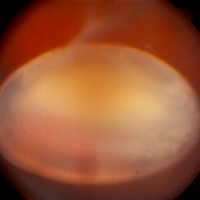
Patient, 65-years-old, male, suffered trauma blunt (the jackpot) in the right eye. Ultrasound of the eye found dislocation the total of the crystalline.
Photographer: JEFFERSON R SOUSA - Institute Dr. Suel Abujamra / São Paulo - Brazil
Imaging device: Topcon TRC-50VT, Film Kodak Ektachrome 160 - ASA 100 / 35mm, field of 35 degrees. Flash 100.
Condition/keywords: dislocated lens, lens luxation
-
 Luxated lens to anterior segment
Sep 7 2022 by JEFFERSON R SOUSA, Tecg.º (Biomedical Systems Technology)
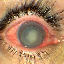
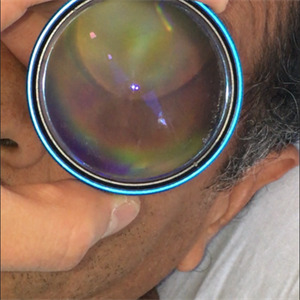
Patient 61 years old, Female, subta low vision after blunt trauma. In the anterior segment photograph, the presence of a lens in the anterior chamber is observed. In the previous follow-up OCT, the disorganization of this follow-up is clear. Above all, the documentation of these cases is essential for future decisions. This patient was urgently referred for a surgical procedure, mainly to control the intraocular pressure, which was at 60 IOP.
Photographer: JEFFERSON ROCHA DE SOUSA - Retinal Department at Instituto Dr. Suel Abujamra Sao Paulo-Brazil.
Imaging device: Optical Coherence Tomography System - OCT CIRRUS 5000, Protocol Wide Angle to Angle.
Condition/keywords: lens luxation, Luxated lens to anterior segment, subluxation of lens
-
 Luxated lens to anterior segment
Sep 7 2022 by JEFFERSON R SOUSA, Tecg.º (Biomedical Systems Technology)
Patient 61 years old, Female, subta low vision after blunt trauma. In the anterior segment photograph, the presence of a lens in the anterior chamber is observed. In the previous follow-up OCT, the disorganization of this follow-up is clear. Above all, the documentation of these cases is essential for future decisions. This patient was urgently referred for a surgical procedure, mainly to control the intraocular pressure, which was at 60 IOP.
Photographer: JEFFERSON ROCHA DE SOUSA - Retinal Department at Instituto Dr. Suel Abujamra Sao Paulo-Brazil.
Imaging device: Clarus 700 - Zeiss,
Condition/keywords: lens luxation, Luxated lens to anterior segment, subluxation of lens
-
 Luxated lens to anterior segment
Sep 7 2022 by JEFFERSON R SOUSA, Tecg.º (Biomedical Systems Technology)
Patient 61 years old, Female, subta low vision after blunt trauma. In the anterior segment photograph, the presence of a lens in the anterior chamber is observed. In the previous follow-up OCT, the disorganization of this follow-up is clear. Above all, the documentation of these cases is essential for future decisions. This patient was urgently referred for a surgical procedure, mainly to control the intraocular pressure, which was at 60 IOP.
Photographer: JEFFERSON ROCHA DE SOUSA - Retinal Department at Instituto Dr. Suel Abujamra Sao Paulo-Brazil.
Imaging device: Optical Coherence Tomography System - OCT CIRRUS 5000, Protocol Wide Angle to Angle.
Condition/keywords: lens luxation, LUX, Luxated lens to anterior segment, subluxation of lens
-
 Luxated Microspherophakic Lens FA
Jul 7 2021 by Linda A Cernichiaro- Espinosa, MD
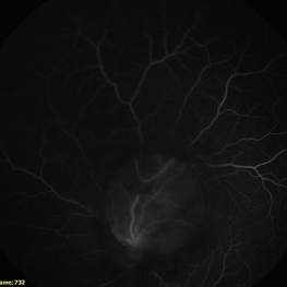
A 12-month-old female with bilateral microspherophakic lens luxation.
Photographer: Linda A Cernichiaro-Espinosa, MD
Condition/keywords: fluorescein angiogram (FA), lens luxation, microspherophakia, pars plana vitrectomy (PPV), pediatric retina
-
 Luxated Microspherophakic Lens FA
Jul 7 2021 by Linda A Cernichiaro- Espinosa, MD
A 12-month-old female with bilateral microspherophakic lens luxation.
Photographer: Linda A Cernichiaro-Espinosa, MD
Condition/keywords: fluorescein angiogram (FA), lens luxation, microspherophakia, pars plana vitrectomy (PPV), pediatric retina
-
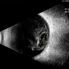 Ocular B-scan Ultrasound (Longitudinal Scan M6, gain 100 dB)
Jun 26 2025 by Hector Gabriel Moreno Solano, MD, MHA
B-scan ultrasound was performed in longitudinal section M6 with a gain of 100 dB. A hyperechoic structure with posterior acoustic shadowing is observed, consistent with lens luxation and condensed vitreous bands adjacent to the lens. The dislocated lens measures approximately 9.54 mm x 4.62 mm. The study was conducted following blunt ocular trauma caused by a golf ball. The remaining vitreous cavity appears anechoic, with no evidence of retinal detachment or other structural abnormalities in this section.
Photographer: Hector Gabriel Moreno Solano, Instituto Mexicano de Oftalmología “IMO I.A.P”
Imaging device: Quantel Medical
Condition/keywords: B scan ultrasound, lens luxation, ocular trauma
-
 Traumatic Luxated Lens to Vitreous Cavity
Sep 11 2018 by PAVEL FLORES-MORENO
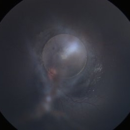
Examination of middle-aged male patient after left eye trauma.
Photographer: Pavel Flores, Centro Médico Nacional Siglo XXI IMSS
Condition/keywords: lens luxation
 Loading…