Search results (241 results)
-
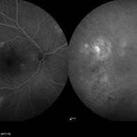
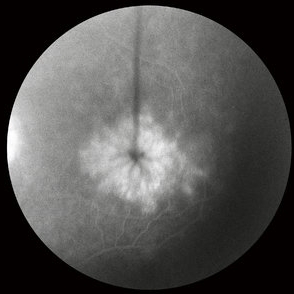
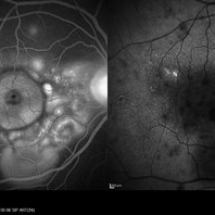
 Acute Multifocal Placoid Pigment Epitheliopathy
Sep 15 2014 by Thomas A. Ciulla, MD, MBA, FASRS
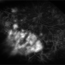
AMPPE in a 42-year-old woman. Late phase angiography shows staining of multiple focal lesions.
Photographer: Thomas Steele
Condition/keywords: acute multifocal placoid pigment epitheliopathy (AMPPE), late phase
-
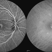
 Central Retinal Artery Occlusion
May 25 2017 by Olivia Rainey
UItra-widefield fluorescein angiography, taken at 6 minutes and 22 seconds, of an 73-year-old woman with a central retinal artery occlusion in her right eye.
Photographer: Olivia Rainey
Imaging device: Optos California
Condition/keywords: central retinal artery occlusion (CRAO), fluorescein angiogram (FA), ischemia, late phase, non-perfusion, Optos, ultra-wide field imaging
-
 Central Serous Chorioretinopathy
Jan 25 2022 by Olivia Rainey
Late phase widefield fluorescein angiography of a 60-year-old male with Central Serous Chorioretinopathy. Chronic history of CSR followed with observation without treatment prior to presenting at our office. The physician noted significant findings on exam and imaging with multifocal areas of inactive and active changes OD. FA shows superotemporal macular leakage, subtle inferonasal macular leakage and staining as well as multifocal hypercyanescence on ICG. Fortunately foveal sparing and thus observation is recommended at this time OD.
Photographer: Olivia Rainey, OCT-C, COA
Imaging device: Heidelberg Spectralis
Condition/keywords: 55-degrees, central serous chorioretinopathy (CSCR), central serous retinopathy (CSR), chronic central serous chorioretinopathy (CSCR), fluorescein angiogram (FA), fluorescein leakage, heidelberg spectralis, indocyanine green (ICG) angiography, late phase
-
 Central Serous Chorioretinopathy with Smokestack Late Phase
Oct 8 2012 by Jeffrey G. Gross, MD, FASRS
CSCR with smokestack, late phase.
Condition/keywords: central serous chorioretinopathy (CSCR), late phase, smokestack
-
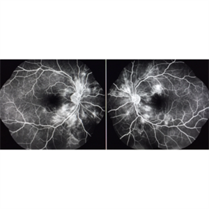
 Central Serous Retinopathy
May 16 2017 by Olivia Rainey
Simultaneous fluorescein and indocyanine green angiography of an 37-year-old male with central serous retinopathy affecting his right eye. Patient's vision declined from 20/25 to 20/80 in the right eye. He elected for treatment with photodynamic therapy.
Photographer: Olivia Rainey
Imaging device: Heidelberg Spectralis
Condition/keywords: 30 degrees, central serous retinopathy (CSR), fluorescein angiogram (FA), fluorescein leakage, Heidelburg Spectralis, indocyanine green (ICG) angiography, late phase, mushroom cloud
-
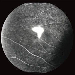
 Choroidal Hemangioma
Sep 15 2014 by Thomas A. Ciulla, MD, MBA, FASRS
Late phase fuorescein angiography reveals a well-circumscribed coarse hyperfluorescent vascular pattern within the lesion. There is no leakage.
Photographer: Charlotte Harris
Condition/keywords: choroidal hemangioma, choroidal tumor, late phase
-
 Cystoid Macular Edema
Oct 8 2012 by Jeffrey G. Gross, MD, FASRS
CME, s/p AC-IOL, FA late phase.
Condition/keywords: cystoid macular edema (CME), late phase
-
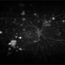
 Familial Drusen with Extrafoveal Pigment Epithelial Detachment
Nov 21 2019 by Olivia Rainey
Late images from a fluorescein angiogram of a 66-year-old female with familial drusen with extrafoveal pigment epithelial detachments affecting both eyes. The physician is presuming that the small multifocal PED's result from her familial drusen. These remain asymptomatic and clinically stable on her exam 11-20-19. VA OD: Dcc20/25-1+1 and VA OS: Dcc20/25-2.
Photographer: Olivia Rainey
Imaging device: Heidelberg Spectralis
Condition/keywords: drusen, drusenoid PED, familial drusen, fluorescein angiogram (FA), hyperfluorescence, late phase, pigment epithelial detachment (PED)
-
 Hypertensive Retinopathy
Nov 14 2019 by Jennifer Schiefer, CRA, COA
46-year-old male who suffered CVA from hypertensive crisis prior to these images. Patient presented in office reporting episode of dim Va OS that happened several days prior. Va was 20/25 OU upon examination.
Photographer: Jennifer Schiefer
Condition/keywords: exudates, fluorescein angiogram (FA), hypertension, hypertensive retinopathy, ischemia, late phase
-
 Proliferative Diabetic Retinopathy
Mar 29 2019 by Olivia Rainey
Ultra-wide field fluorescrein angiogram of a 48-year-old male with proliferative diabetic retinopathy affecting his right eye. Patient was treated with pan-retinal laser follow his fluorescein angiogram and will likely need intravitreal injections in the near future.
Photographer: Olivia Rainey
Imaging device: Optos
Condition/keywords: diabetes, diabetic macular edema, fluorescein angiogram (FA), fluorescein leakage, late phase, Optos, proliferative diabetic retinopathy (PDR), ultra-wide field imaging
-
 Proliferative Diabetic Retinopathy with Active Neovascularization
Jul 30 2019 by Olivia Rainey
Ultra-wide field fluorescein angiogram of a 36-year-old male with proliferative diabetic retinopathy with active neovascularization affecting his left eye. Patient presented with seeing flashing lights and trouble seeing to drive at night. His vision was sc20/50-1 PH20/40-2 in the left eye. There are suspicious vessels within the inferonasal retina of the patient's left eye. Labs ordered and are negative for sickle cell.
Photographer: Olivia Rainey
Imaging device: Optos
Condition/keywords: diabetes, diabetic macular edema, fluorescein angiogram (FA), fluorescein leakage, ischemia, late phase, left eye, neovascularization (NV), Optos, proliferative diabetic retinopathy (PDR), ultra-wide field imaging
-
 Proliferative Diabetic Retinopathy with Retinal Ischemia
Mar 29 2019 by Olivia Rainey
Ultra-wide field fluorescrein angiogram of a 42-year-old female with proliferative diabetic retinopathy with retinal ischemia affecting her right eye. Patient had been noticing a vision decline and floaters over the past few months. She was treated with Avastin in her right eye and then her left one week later.
Photographer: Olivia Rainey
Imaging device: Optos
Condition/keywords: diabetes, diabetic macular edema, fluorescein angiogram (FA), fluorescein leakage, late phase, neovascularization of the disc (NVD), Optos, proliferative diabetic retinopathy (PDR), retinal ischemia, ultra-wide field imaging
-
 Retinal Cavernous Hemangioma
Oct 22 2020 by Olivia Rainey
Ultra-widefield fluorescein and ICG angiogram of a 31-year-old male presenting with a retinal cavernous hemangioma affecting his left eye. Patient was 18-years-old when he was diagnosed with a retinal cavernous hemangioma. He has had a few episodes of vitreous hemorrhages since then. His vision was 20/20-1 in both eyes.
Photographer: Becca Harris
Imaging device: Optos California
Condition/keywords: cavernous hemangioma of the retina, fluorescein angiogram (FA), indocyanine green (ICG) angiography, late phase, left eye, Optos, ultra-wide field imaging
-
 Syphilis Neuroretinopathy
Sep 25 2013 by Alexandre Durao Alves Pereira, MD
Syphilis neuroretinopathy late phase FA.
Photographer: Alexandre Pereira
Condition/keywords: late phase
-
 Syphilis Neuroretinopathy (Color Photo)
Sep 25 2013 by Alexandre Durao Alves Pereira, MD
Syphilis neuroretinopathy, late phase FA, (color photograph).
Photographer: Alexandre Pereira
Condition/keywords: color photo, late phase, syphilis neuroretinopathy
-
 Syphilis Neuroretinopathy (Late Phase FA)
Sep 25 2013 by Alexandre Durao Alves Pereira, MD
Syphilis neuroretinopathy, (late phase FA).
Photographer: Alexandre Pereira
Condition/keywords: late phase
-
 Syphilis Neuroretinopathy (Late Phase FA)
Sep 25 2013 by Alexandre Durao Alves Pereira, MD
Syphilis neuroretinopathy, (late phase FA).
Photographer: Alexandre Pereira
Condition/keywords: late phase
-
 Syphilis Neuroretinopathy (Red Free Photo)
Sep 25 2013 by Alexandre Durao Alves Pereira, MD
Syphilis neuroretinopathy, late phase FA, (red free photograph).
Photographer: Alexandre Pereira
Condition/keywords: late phase, red-free
-
 Syphilitic Uveitis
Apr 2 2020 by Olivia Rainey
Ultrawide-field fluorescein angiogram of a 42-year-old male with syphilitic uveitis affecting his right eye more than his left. Patient is HIV positive. He developed hearing loss and palm/leg/scalp rash prompting diagnosis of neurosyphilis, s/p IM and full IV course of 2.4 Mil PCN G, and finished this course 3/9/20. He admits to recent rectal bleeding with ongoing plan for colonoscopy 3/16/20. He has a history of extensive travel including London, Hong Kong, and Bangkok. His husband has also been treated with IV PCN G, however per chart review he has multiple sexual partners. Patient's vision was 20/20 in each eye.
Photographer: Olivia Rainey
Imaging device: Optos California
Condition/keywords: disc hyperfluorescence, FA late phase leakage, fluorescein angiogram (FA), fluorescein leakage, HIV, late phase, optic nerve edema, Optos, phelbitis, syphilis neuroretinopathy, ultra-wide field imaging, uveitis
-
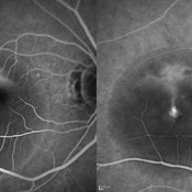
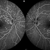
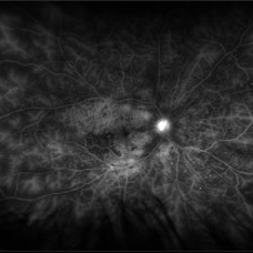
 Vogt Koyanagi Harada
Oct 7 2015 by Avris Romario Diparaja Siahaan
Simultaneous FA + ICG (Late Phase) of a 42-year-old woman with Harada Syndrome in both eyes.
Photographer: Yohanes Harry Purwanto, Klinik Mata Nusantara
Imaging device: Heidelberg HRA + OCT
Condition/keywords: indocyanine green (ICG) angiography, late phase, Vogt-Koyanagi-Harada
-
 Acute Central Serous Chorioretinopathy
Sep 15 2012 by Hamid Ahmadieh, MD
Late phase FA & ICG angiography images of a 30-year-old man with acute CSCR.
Photographer: Hamid Ahmadieh, MD, Ophthalmic Research Center, Labbafinejad Medical Center, Shahid Beheshti University of Medical Sciences
Imaging device: Heidelberg HRA
Condition/keywords: central serous chorioretinopathy (CSCR), indocyanine green (ICG) angiography
-
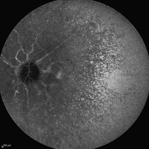
 Acute Posterior Multifocal Placoid Pigment Epitheliopathy
Sep 15 2012 by Roy D. Brod, MD
Late phase fluorescein angiogram demonstrating staining of placoid lesions in patient with APMPPE.
Photographer: Julia Walker
Condition/keywords: acute posterior multifocal placoid pigment epitheliopathy (APMPPE)
-
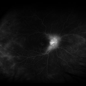
 Angioid Streaks
Sep 29 2012 by Hamid Ahmadieh, MD
Late phase ICG angiography image of the right eye of a 59-year-old man with angioid streaks.
Photographer: Hamid Ahmadieh, MD; Ophthalmic Research Center, Labbafinejad Medical Center, Shahid Beheshti University of Medical Sciences
Imaging device: Heidelberg Spectralis
Condition/keywords: angioid streaks, indocyanine green (ICG) angiography
-
 Angioid Streaks
Sep 29 2012 by Hamid Ahmadieh, MD
Late phase ICG angiography image of the left eye of a 59-year-old man with angioid streaks.
Photographer: Hamid Ahmadieh, MD; Ophthalmic Research Center, Labbafinejad Medical Center, Shahid Beheshti University of Medical Sciences
Imaging device: Heidelberg Spectralis
Condition/keywords: angioid streaks, indocyanine green (ICG) angiography
-
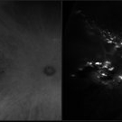
 Angioid Streaks & CNV (Fig 4)
Aug 25 2012 by Hamid Ahmadieh, MD
Late phase ICG angiography imaging of a 53-year-old woman with a juxtafoveal CNV secondary to angioid streaks.
Photographer: Hamid Ahmadieh, Ophthalmic Research Center, Labbafinejad Medical Center
Imaging device: Heidelberg Spectralis
 Loading…