Search results (12 results)
-
 Busacca nodules
May 2 2013 by Henry J. Kaplan, MD
Typical Busacca iris stromal nodules in sarcoid uveitis; notice the ps formation.
Condition/keywords: busacca nodulaes, granulomatous uveitis, iris nodules, sarcoid bussaca iris nodules
-
 Granulomatous Uveitis
May 18 2020 by McGill University Health Centre
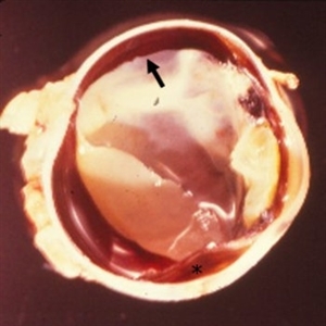
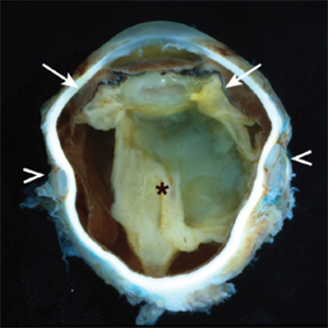
Granulomatous uveitis is found in many inflammatory diseases, and is generally characterized by a predominant histiocytic infiltrate forming a “wall” (granuloma) around a pathogen or foreign body. This is an example of granulomatous uveitis. The eye is aphakic; the uveal track is thickened; and a granuloma is present and attached to the endothelium of the cornea (arrow). The anterior chamber is filled with a hazy material (arrowhead). The vitreous is fibrotic and tractional bands are also present (*).
Condition/keywords: granulomatous uveitis
-
 VKH Pseudotumor – Chronic Subretinal Fibrosis
May 11 2025 by Felipe Murati
Ultra-widefield fundus image from a 36-year-old woman with chronic VKH syndrome showing a pseudotumor-like subretinal fibrotic lesion in the right eye. The lesion developed after multiple relapses and remained stable over a 1-year follow-up with immunosuppressive treatment including prednisone, mycophenolate mofetil, and adalimumab. No active choroiditis or exudative detachment was observed. Multimodal imaging was essential for disease monitoring.
Photographer: Felipe A. Murati, MD, University of Arizona
Imaging device: Optos California ultra-widefield retinal imaging system, single-capture, color fundus modality.
Condition/keywords: adalimumab, chronic inflammation, granulomatous uveitis, OCT, Optos ultra-widefield imaging, pseudotumor, subretinal fibrosis, VKH, Vogt-Koyanagi-Harada
-
 VKH Pseudotumor – Fluorescein Angiography
May 11 2025 by Felipe Murati
Fluorescein angiography image from a 36-year-old woman with chronic Vogt-Koyanagi-Harada (VKH) syndrome showing a pseudotumor-like lesion with late-phase staining and no active leakage. The image highlights subretinal fibrosis in the right eye, stable under long-term immunosuppressive therapy with mycophenolate mofetil and adalimumab. No signs of active choroiditis are present, confirming a quiescent phase.
Photographer: Felipe A. Murati, MD, University of Arizona
Imaging device: Optos California, fluorescein angiography modality
Condition/keywords: choroiditis, Fluorescein angiography, granulomatous uveitis, Optos FA, pseudotumor, subretinal fibrosis, VKH, Vogt-Koyanagi-Harada
-
 Nongranulomatous Uveitis
May 18 2020 by McGill University Health Centre
Nongranulomatous uveitis is a group of diseases often related to systemic autoimmune diseases such as arthritis. In this enucleation specimen, the choroid is diffusely thickened (arrow) and a choroidal detachment is present (*). The retina is opaque and the lens is cataractous.
Condition/keywords: uveitis
-
 Nongranulomatous Uveitis
May 18 2020 by McGill University Health Centre
Nongranulomatous uveitis is a group of diseases often related to systemic autoimmune diseases such as arthritis. This image shows a thickened uveal track (arrows) and a complete, long-standing retinal detachment (*). The lens is cataractous and the iris, ciliary body, and parts of the retina are attached to it. A scleral buckle is present (arrowheads).
Condition/keywords: uveitis
-
 Ocular Tuberculosis
May 18 2020 by McGill University Health Centre
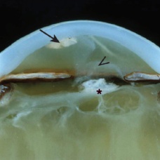
Ocular tuberculosis refers to necrotizing granulomatous uveitis caused by Mycobacterium tuberculosis infection. The mechanism of infection of ocular structures is via hematogenous dissemination of the bacteria during the primary infection. In this enucleation specimen, the anterior chamber and vitreous cavity are filled by caseous exudate. The iris is also replaced by a whitish material, and the retina is completely detached. Note the whitish thickening of the posterior choroid (arrow).
Condition/keywords: caseous exudate, enucleation, ocular tuberculosis, uveitis
-
 Sarcoidosis Panuveitis Slide 3
Oct 22 2012 by Ronald C. Gentile, MD
Gonioscopic photograph reveals peripheral anterior synechiae with granuloma involving the iris root and Bussaca nodules on the iris surface consistent with granulomatous uveitis.
Photographer: The New York Eye & Ear Infirmary Department of Medical Imaging
Condition/keywords: sarcoid granuloma, sarcoidosis panuveitis
-
 Sarcoidosis Panuveitis Slide 4
Oct 22 2012 by Ronald C. Gentile, MD
High frequency ultrasound biomicroscopy of the anterior chamber and angle images a granuloma involving the iris root and Bussaca nodules on the iris surface consistent with granulomatous uveitis.
Photographer: The New York Eye & Ear Infirmary Department of Medical Imaging
Condition/keywords: sarcoid granuloma, sarcoidosis panuveitis
-
 Slide 2-39
Feb 19 2019 by Lancaster Course in Ophthalmology
Cystic macula in chronic nongranulomatous uveitis.
Condition/keywords: chronic nongranulomatous uveitis, cystoid macular degeneration
-
 Slide 2-40
Feb 19 2019 by Lancaster Course in Ophthalmology
End-stage of chronic nongranulomatous uveitis in a 47-year-old male with arthritis, showing iris bombe, cystic separation of the iris pigment layers, intumescent cataract, cyclitic membrane, fresh hemorrhage from new oral vessels, and complete retinal detachment.
Condition/keywords: cataract, chronic nongranulomatous uveitis, cyclitic membrane, iris bombe
-
 Sympathetic Ophthalmia
May 18 2020 by McGill University Health Centre
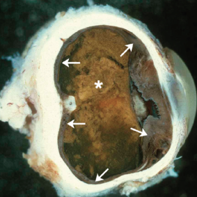
Sympathetic ophthalmia is characterized by bilateral diffuse granulomatous uveitis that occurs 2 weeks to many years after traumatic penetration or perforation of the eye. It threatens the sight of the uninjured (sympathizing) eye. In this enucleation specimen, thickening of the uveal tract is evident (arrows). Complete proteinaceous retinal detachment (*) is also present, along with posterior synechia (adhesion of the iris to the anterior capsule of the lens).
Condition/keywords: enucleation, sympathetic ophthalmia
 Loading…