Search results (189 results)
-
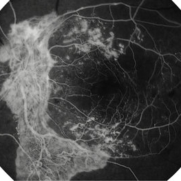
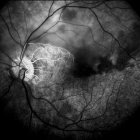
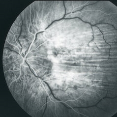
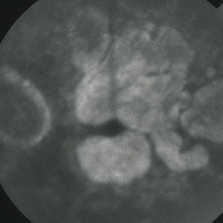
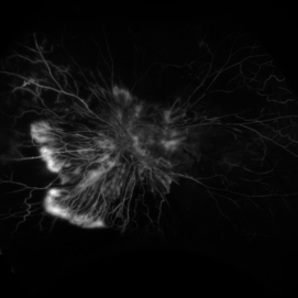 Active Proliferative Diabetic Retinopathy
Aug 16 2022 by Donnie Willis
51 yo female. Uncontrolled Diabetes. Active Proliferative Diabetic Retinopathy.
Photographer: Donnie Willis, Tennessee Retina
Imaging device: Optos
Condition/keywords: capillary dropouts, Diabetes, fluorescein angiogram (FA), OPTOS, proliferative diabetic retinopathy (PDR), tractional retinal detachment
-
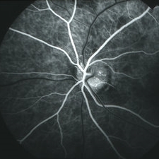
 Acute Central Retinal Artery Occlusion
Jul 27 2022 by Becca Harris
Ultra widefield FA/ICG of a 24 year old female with an acute central retinal artery occlusion affecting the right eye. Patient presented with extreme headaches following DAVF surgery the previous day. Patient has Factor VIII deficiency and had a cerebral venous thrombosis 9 years ago and lost vision in the right eye at that time. Patient has history of optic sheath fenestration OU and craniotomy. On initial evaluation, she had a CRAO as well as diffuse choroidal nonperfusion noted on optos FA. Suspect nonperfusion to third and sixth nerve leading to palsy. Occlusion of vasculature in the setting of recent endovascular embolization of fistulas in the CNS. Discussed diagnosis and poor prognosis with parents and patient. Patient had no light perception at the time of her initial appointment.
Photographer: Becca Harris
Imaging device: Optos California
Condition/keywords: Choroidal non-perfusion, fluorescein angiogram (FA), indocyanine green (ICG) angiography, non-perfusion, Optos, Right Eye, ultra-wide field imaging
-
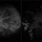
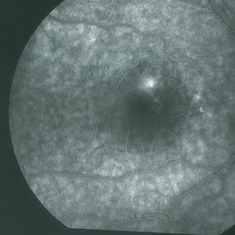
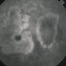
 Acute Posterior Multifocal Placoid Pigment Epitheliopathy
Feb 20 2024 by Soobien Lee
Fluorescein angiogram of a 20-year-old caucasian female with viral prodrome and vision loss OS>OD secondary to Acute Posterior Multifocal Placoid Pigment Epitheliopathy (APPME). Early blockage with late hyperfluorescent leakage can be seen on fluorescein angiography of the left eye.
Photographer: Ashley Metzger, Elman Retina Group
Imaging device: Optos Ultra-Widefield Fluorescein Angiography
Condition/keywords: acute posterior multifocal placoid pigment epitheliopathy (APMPPE), bacilliary layer detachment, FA, FA early phase, fluorescein angiogram (FA), Optos, uveitis, white dot syndrome
-
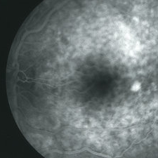
 Acute Posterior Multifocal Placoid Pigment Epitheliopathy
Feb 20 2024 by Soobien Lee
Fluorescein angiogram of a 20-year-old caucasian female with viral prodrome and vision loss OS>OD secondary to Acute Posterior Multifocal Placoid Pigment Epitheliopathy (APPME). Early blockage with late hyperfluorescent leakage can be seen on fluorescein angiography of the left eye.
Photographer: Ashley Metzger, Elman Retina Group
Imaging device: Optos Ultra-Widefield Fluorescein Angiography
Condition/keywords: acute posterior multifocal placoid pigment epitheliopathy (APMPPE), bacilliary layer detachment, FA, FA late phase, FA late phase leakage, fluorescein angiogram (FA), Optos, uveitis, white dot syndrome
-
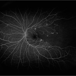
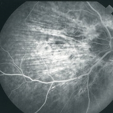
 Acute Zonal Occult Outer Retinopathy (AZOOR) FA, Fluorescein Angiography, Peripheral Vasculitis
Jan 19 2022 by James B. Soque, CRA, OCT-C, COA, FOPS
Acute Zonal Occult Outer Retinopathy (AZOOR). Peripheral Vasculitis OD. Fluorescein angiography showing vasculitis in the far right periphery 8-10 o'clock. 46-year-old white male, VA CC 20/16, 20/12.5, has had recurrent vasculitis for 11 years. No treatment.
Photographer: James Soque, CRA, OCT-C, COA, FOPS, Island Retina, Shirley, NY
Imaging device: Optos California
Condition/keywords: acute zonal occult outer retinopathy (AZOOR), FA early phase, fluorescein angiogram (FA), Peripheral Vasculitis, ultra-wide field imaging
-
 Acute Zonal Occult Outer Retinopathy (AZOOR) FA, Ultra Wide-Field Fluorescein Angiogram Early
Jan 19 2022 by James B. Soque, CRA, OCT-C, COA, FOPS
Acute Zonal Occult Outer Retinopathy, FA, Fluorescein Angiography, OD. 46-year-old white male, VA CC 10/16, 20/12.5, has had recurrent vasculitis for 11 years. No treatment.
Photographer: James Soque, CRA, OCT-C, COA, FOPS, Island Retina, Shirley, NY
Imaging device: Optos California
Condition/keywords: acute zonal occult outer retinopathy (AZOOR), FA EARLY, fluorescein angiogram (FA), ultra-wide field imaging
-
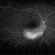
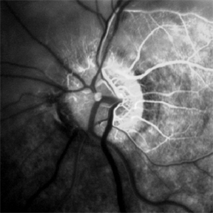
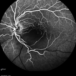
 Advanced Proliferative Diabetic Retinopathy With Fibrovascular Proliferation
Jan 4 2019 by Isha Agarwalla
A 29-year-old female with a long-standing history of diabetes mellitus presented with a fibrovascular membrane(FVM) at the viteroretinal interface due to underlying inflammation and angiogenesis induced by ischemia. FVM involved the disc and extended towards the superior and inferior arcades along with extensive capillary drop out areas due to micro aneurysms.
Condition/keywords: fibrovascular proliferation, fluorescein angiogram (FA), proliferative diabetic retinopathy (PDR)
-
 Atypical Arterial Beading Secondary to Retinal Capillary Hemangioma
Jan 28 2020 by Sophia El Hamichi, MD
Image 1: Fundus picture montage depicting RCH with feeder and drainer vessels. Note the unusual beaded appearance of the arterioles. Image 2: 2A: Arterial phase of fluorescein angiography shows early filling of the arteriole. 2B: Arterio-venous phase highlighting the sausage appearance of the arterioles beading.
Condition/keywords: arterial beading, fluorescein angiogram (FA), retinal capillary hemangioblastoma, retinal capillary hemangioma
-
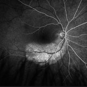
 Best Disease
Nov 7 2024 by Virginia Gebhart
Fluorescein angiogram of 49 year female with Best Disease. Genetic testing done in 2000 confirms Best Disease and also possible Stargardts mutation. Characteristic bullseye maculopathy with surrounding yellowish flecks are present in both eyes.
Photographer: Virginia Gebhart, Retina Consultants of Carolina
Imaging device: Optos California
Condition/keywords: Best disease, fluorescein angiogram (FA)
-
 Bilateral Central Serous Retinopathy
Mar 26 2019 by Gary R. Cook, MD, FACS
Late-phase fluorescein angiogram image of the right eye of a 37-year-old white male showing pinpoint leak with late diffusion of dye from it superiorly and RPE irregularities nasal to fovea in a case of bilateral central serous retinopathy; VA = 20/20-2.
Imaging device: Topcon VT-50
Condition/keywords: central serous retinopathy (CSR), FA late phase, fluorescein angiogram (FA)
-
 Bilateral Central Serous Retinopathy
Mar 26 2019 by Gary R. Cook, MD, FACS
Mid-phase fluorescein angiogram frame of a pinpoint leak in the temporal macula OS of a 37-year-old white male with bilateral central serous retinopathy; VA = 20/15+3.
Imaging device: Topcon VT-50
Condition/keywords: central serous retinopathy (CSR), FA mid phase, fluorescein angiogram (FA)
-
 Branch Retinal Artery Occlusion
Sep 11 2018 by Olivia Rainey
Ultra-wide field fluorescein angiogram of a 46-year-old male with a branch retinal artery occlusion affecting his left eye. The longstanding occlusion and has resulted in peripheral nonperfusion and neovascularization.
Photographer: Olivia Rainey
Imaging device: Optos
Condition/keywords: branch retinal artery occlusion (BRAO), fluorescein angiogram (FA), left eye, neovascularization (NV), non-perfusion, Optos
-
 Branch Retinal Artery Occlusion With Calcium Embolus at the Disc - Fundus Fluorescence Angiogram (FA)
Apr 7 2018 by Rameez N Hussain, MD
Acute branch retinal artery occlusion with a calcium embolus at the disc which is hyper-fluorescent in FA.
Photographer: DR RAMEEZ N HUSSAIN
Imaging device: ZEISS
Condition/keywords: branch retinal artery occlusion (BRAO), embolus, fluorescein angiogram (FA), retinal edema
-
 Branch Retinal Artery Occlusion With Calcium Embolus at the Disc - Fundus Fluorescence Angiogram (FA)
Apr 7 2018 by Rameez N Hussain, MD
Acute branch retinal artery occlusion with a calcium embolus at the disc which is hyperfluorescent in FA.
Photographer: DR RAMEEZ N HUSSAIN
Imaging device: Zeiss
Condition/keywords: branch retinal artery occlusion (BRAO), embolus, fluorescein angiogram (FA), retinal edema
-
 Branch Retinal Vein Occlusion
Aug 22 2024 by Virginia Gebhart
Fluorescein angiogram of branch retinal vein occlusion in 75 year old female. Scattered microaneurysms with late CME and persistent SRF. Pt will consider laser treatment but is hesitant for injections at this time due to possible side effects.
Photographer: Virginia Gebhart
Imaging device: Optos California
Condition/keywords: branch retinal vein occlusion (BRVO), BRVO, cystoid macular edema (CME), FA, FA late phase, fluorescein angiogram (FA), macular edema, microaneurysms, retinal microaneurysms
-
 Branch Retinal Vein Occlusion With Peripheral Pigmentary Change
Jan 15 2019 by Olivia Rainey
Ultra-wide field fluorescein angiogram of an 85-year-old female with a branch retinal vein occlusion with peripheral pigmentary changes. Patient developed a BRVO after a PPV for an epiretinal membrane.
Photographer: Olivia Rainey
Imaging device: Optos
Condition/keywords: branch retinal vein occlusion (BRVO), epiretinal membrane (ERM), fluorescein angiogram (FA), left eye, Optos, pigmentary retinal dystrophy
-
 C-R Folds
Mar 26 2019 by Gary R. Cook, MD, FACS
Mid-phase FA image of the right eye of a white male with bilateral C-R folds showing alternating hyper- and hypofluorescent bands.
Imaging device: Topcon VT-50
Condition/keywords: bilateral chorioretinal folds, chorioretinal fold, FA mid phase, fluorescein angiogram (FA)
-
 C-R Folds
Mar 26 2019 by Gary R. Cook, MD, FACS
Early phase FA frame of the left eye of a WM with bilateral C-R folds showing alternating hyper- and hypofluorescent bands.
Imaging device: Topcon VT-50
Condition/keywords: bilateral chorioretinal folds, chorioretinal fold, FA early phase, fluorescein angiogram (FA)
-
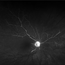
 Capillary Hemangioma
Apr 1 2019 by Gary R. Cook, MD, FACS
Mid-phase (laminar venous phase) fluorescein angiogram image of a capillary hemangioma of the optic disc OS showing delayed filling and relative hypofluorescence in the area of the hemangioma on the superior aspect in a 28-year-old white female
Imaging device: Topcon VT-50
Condition/keywords: FA mid phase, fluorescein angiogram (FA), hemangioma
-
 Capillary Hemangioma
Apr 1 2019 by Gary R. Cook, MD, FACS
Late-phase (6 minutes) fluorescein angiogram image of a capillary hemangioma of the optic disc OS in a 28-year-old, asymptomatic white female showing late staining in the area of the hemangioma superiorly.
Imaging device: Topcon VT-50
Condition/keywords: FA late phase, fluorescein angiogram (FA), hemangioma, retinal capillary hemangioma
-
 Central Areolar Choriocapillaris Atrophy
Mar 26 2019 by Gary R. Cook, MD, FACS
Late-phase fluorescein angiogram image of the right eye of a 64-year-old white male with central areolar choriocapillaris atrophy showing late leakage from intact choriocapillaris around the perimeter of the disc and macular areas of choriocapillaris atrophy; VA= 20/50
Imaging device: Topcon VT-50
Condition/keywords: FA late phase, fluorescein angiogram (FA), hereditary choroidal atrophy, hereditary choroidal dystrophy
-
 Central Areolar Choriocapillaris Atrophy
Mar 26 2019 by Gary R. Cook, MD, FACS
Late-phase fluorescein angiogram image of the left eye of a 64-year-old white male with central areolar choriocapillaris atrophy showing light late staining of the central lesions OS; V.A. = 20/30
Imaging device: Topcon VT-50
Condition/keywords: choriocapillaris, FA late phase, fluorescein angiogram (FA), hereditary choroidal atrophy, hereditary choroidal dystrophy
-
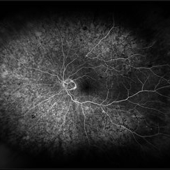
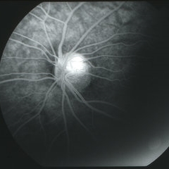
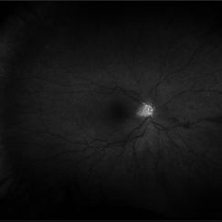
 Central Retinal Artery Occlusion
May 16 2017 by Olivia Rainey
Fluorescein angiogram of an 66-year-old female with a central retinal artery occlusion affecting her left eye.
Photographer: Olivia Rainey
Imaging device: Heidelberg Spectralis
Condition/keywords: 50 degrees, central retinal artery occlusion (CRAO), fluorescein angiogram (FA), left eye, mid phase, retinal ischemia
-
 Central Retinal Artery Occlusion
May 25 2017 by Olivia Rainey
Ultra-wide field fluorescein angiography, taken at 42 seconds, of an 73-year-old female with a central retinal artery occlusion in her right eye.
Photographer: Olivia Rainey
Imaging device: Optos California
Condition/keywords: central retinal artery occlusion (CRAO), early phase, fluorescein angiogram (FA), ischemia, non-perfusion, Optos, ultra-wide field imaging
-
 Central Retinal Artery Occlusion
May 25 2017 by Olivia Rainey
UItra-widefield fluorescein angiography, taken at 6 minutes and 22 seconds, of an 73-year-old woman with a central retinal artery occlusion in her right eye.
Photographer: Olivia Rainey
Imaging device: Optos California
Condition/keywords: central retinal artery occlusion (CRAO), fluorescein angiogram (FA), ischemia, late phase, non-perfusion, Optos, ultra-wide field imaging
 Loading…