Search results (5 results)
-
 Slide 12-18
Feb 27 2019 by Lancaster Course in Ophthalmology
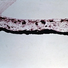
Rubeosis iridis. Fibrovascular tissue is present abnormally anterior to the anterior border layer of the iris. Shrinkage of such abnormal tissue results frequently in ectropion uveae (H&E xlOl).
Condition/keywords: fibrovascular tissue, rubeosis
-
 Slide 8-4
Mar 4 2019 by Lancaster Course in Ophthalmology
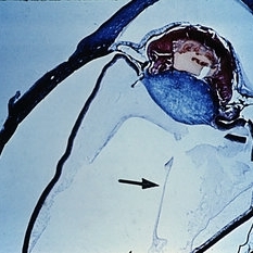
Persistence and hyperplasia of the primary vitreous (PHPV). The dense fibrovascular tissue molds the posterior surface of the lens and has drawn the ciliary processes and peripheral retina toward the center of the mass. A section of the hyaloid artery is present in the vitreous cavity (arrow). (A.F.l.P. No. 744398)
Condition/keywords: ciliary, fibrovascular tissue, hyaloid artery, persistent hyperplastic primary vitreous (PHPV)
-
 Ultra-Widefield Image of Tractional-Rhegmatogenous Retinal Detachment Sparing Fovea
Jul 16 2021 by Kushal S Delhiwala, MBBS, MS, FMRF,FICO, FAICO
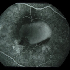
Ultra-widefield fundus photograph of an 45-year-old phakic male with superior tractional-rhegmatogenous retinal detachment sparing fovea. Retinal break was observed at the base of fibrous proliferation. Scattered whitish outer retinal spots were noted in area of retinal detachment.
Photographer: Kushal Delhiwala, Netralaya superspeciality eye hospital, Ahmedabad, Gujarat,India
Imaging device: Optos Daytona
Condition/keywords: fibrovascular proliferation, fibrovascular tissue, outer retinal white spots, tractional retinal detachment, ultra-wide field imaging
-
 Fibrovascular PED
Jun 4 2014 by Henry J. Kaplan, MD
Fluorescein angiography in AV phase demonstrates filling of the PED with an inferonasal notch secondary to the fibrovascular tissue. #2
Condition/keywords: pigment epithelial detachment (PED), vascularized pigment epithelial detachment (PED)
-
 Slide 12-25
Feb 27 2019 by Lancaster Course in Ophthalmology
Sequelae. The anterior chamber is deep, and the pupillary iris shows ectropion uveae resulting from shrinkage of new fibrovascular tissue (rubeosis iridis) on the anterior surface of the iris. The lens is dislocated posteriorly into the vitreous. All changes are the result of blunt trauma.
Condition/keywords: ectropion uveae, rubeosis, sequelae
 Loading…