Search results (5 results)
-
 Slide 1-27
Feb 19 2019 by Lancaster Course in Ophthalmology
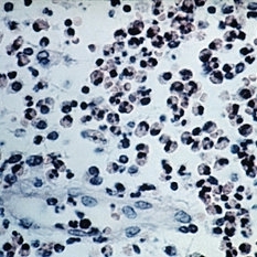
Acute arteritis in an orbital inflammatory pseudotumor. Arterial wall is edematous and thickened with PMNs, lymphocytes, and macrophages. Eosinophils are seen in the tissue at right. (H&E stain)
Condition/keywords: arteritis, edematous, eosinophils, polymorphonuclear leukocytes (PMNs), pseudotumor
-
 Slide 1-7
Feb 19 2019 by Lancaster Course in Ophthalmology
Upper: Bilobed eosinophils. Lower: Large mast cell identified by its purple granules. Both views are from scrapings in allergic conjunctivitis. (Giemsa stain)
Condition/keywords: cell, conjunctivitis, eosinophils
-
 Slide 6-58
Mar 20 2019 by Lancaster Course in Ophthalmology
Eosinophilic granuloma. Infiltration of orbital bone by eosinophils and large histiocytes (H&E x400).
Condition/keywords: eosinophilic, eosinophils, histiocytes
-
 Slide 1-24
Feb 19 2019 by Lancaster Course in Ophthalmology
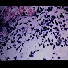
Perivascular eosinophils and PMNs in an acute conjunctivitis. (Giemsa stain)
Condition/keywords: acute conjunctivitis, perivascular eosinophils, polymorphonuclear leukocytes (PMNs)
-
 Slide 8-24
Mar 4 2019 by Lancaster Course in Ophthalmology
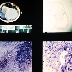
Diffuse nematode endophthalmitis with a fibroinflammatory mass in the vitreous (upper views) with eosinophils and the nematode larva, and total retinal detachment. (E.P. No. 29566)
Condition/keywords: endophthalmitis, fibroinflammatory mass
 Loading…