Search results (9 results)
-
 Slide 4-8
Feb 20 2019 by Lancaster Course in Ophthalmology
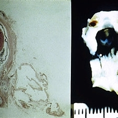
Trisomy 13-15, or nondysjunction (x16). Note the retinal dysplasia and cartilage within the eye. Numerous other anomalies usually are found in babies with this chromosomal aberration. {Scheie Eye Institute, No. 6328.)
Condition/keywords: chromosomal aberration, dysplasia, nondysjunction, Trisomy 13-15
-
 Slide 5-3
Feb 20 2019 by Lancaster Course in Ophthalmology
Loss of orderly maturation of epidermal cells (dysplasia). The red blood cell-like ellipse in the center of the epithelium is a cell which has keratinized out of place (dyskeratosis).
Condition/keywords: dyskeratosis, dysplasia, epidermal cells, epithelium
-
 Slide 6-1
Feb 25 2019 by Lancaster Course in Ophthalmology
Microphthalmos with cyst. Left side shows the macroscopic appearance and right side the microscopic appearance, of a microphthalmic eye with continuou cyst. The eye has multiple anomalies such as hypoplasia of the iris, cataract, nonattachment of the retina, and retinal dysplasia (right, H&E x 1).
Condition/keywords: cataract, cyst, dysplasia, hypoplasia, microphthalmos
-
 Slide 9-4
Feb 25 2019 by Lancaster Course in Ophthalmology
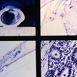
Retinal dysplasia in the Petau syndrome. The microphthalmic eye has a dysplasic retina behind the lens, extending as a strand (arrow) to attach to the retina posteriorly (views on left). The rosettes (asterisks) have a well-developed external limiting membrane (arrow) and photoreceptor outer and inner segments (lower right). There is also a hyperplasic primary vitreous (upper right) with a small nodule of cartilage (arrow).
Condition/keywords: dysplasia, microphthalmos, Patau syndrome, vitreous
-
 Morning Glory
Jun 17 2019 by Feyene Art
Oil on canvas painting inspired by a fundus photograph posted by Alex P. Hunyor, MD. https://imagebank.asrs.org/file/3048/optic-disc-dysplasia
Photographer: Feyene Art
Imaging device: Oil on Canvas Painting
Condition/keywords: Morning Glory Syndrome, optic disc dysplasia
-
 Morning Glory Disc Anomaly
Aug 19 2017 by Mitzy E Torres Soriano, MD
A 10-year-old female patient with morning glory disc anomaly in her left eye.
Photographer: Mitzy E. Torres Soriano
Condition/keywords: coloboma of optic disc, coloboma of the optic nerve, Morning Glory Syndrome, optic disc, optic disc dysplasia
-
 Morning glory optic disc
Jan 11 2013 by Alex P. Hunyor, MD
Morning glory optic disc anomaly, right eye.
Condition/keywords: Morning Glory Syndrome, optic disc dysplasia
-
 Optic disc dysplasia
Jan 11 2013 by Alex P. Hunyor, MD
Optic disc dysplasia - morning glory variant.
Condition/keywords: Morning Glory Syndrome, optic disc dysplasia
-
 Traumatic Giant Retinal Tear Associated Retinal Detachment
Nov 9 2019 by Luis J Haddock, MD
This wide field fundus photograph of the left eye shows a traumatic giant retinal tear associated with total retinal detachment. The image shows the torn superior retina folded over the macula with the underside of the retina visible. There is associated peripheral choroidal detachment due to hypotony from giant retinal tear. This patient has history of spondyloepithelial dysplasia with dwarfism and presented with vision loss after a recent blunt trauma with elbow to the eye.
Imaging device: Optos
Condition/keywords: giant retinal tear, traumatic optic neuropathy
 Loading…