Search results (1584 results)
-
10 steps to follow in PDR
Sep 5 2024 by DAVID PÉREZ GONZÁLEZ, MD
Cases of proliferative diabetic retinopathy with fibrovascular membranes are among the most complex in retinal surgery. Here are 10 steps I follow in each case to achieve the best possible outcome, particularly when performing one-handed dissection with the vitrector.
Condition/keywords: diabetic retinopathy, Fibrovascular, Membranes, Retinal Surgery
-
 Active diabetic retinopathy despite PRP
Oct 30 2022 by Diego Andrés Rodriguez, MD
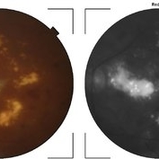
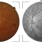
A 52-year-old patient with active proliferative diabetic retinopathy despite good glycemic control and PRP performed 1 year ago in the right eye
Photographer: Sociedad de Cirugía Ocular
Imaging device: Clarus 700
Condition/keywords: diabetic retinopathy, pan-retinal photocoagulation (PRP), proliferative diabetic retinopathy (PDR), wide angle imaging
-
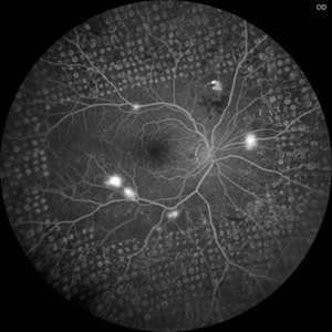
 Active neovascularization in Proliferative Diabetic Retinopathy
Jan 10 2018 by Peter H. Tang, MD, PhD
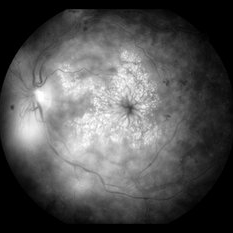
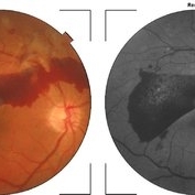
Fluorescein angiography image from a 46-year-old woman with uncontrolled proliferative diabetic retinopathy shows extensive dye leakage from active neovascularization.
Imaging device: Optos California
Condition/keywords: diabetes, diabetic retinopathy, fluorescein leakage, neovascularization elsewhere (NVE), neovascularization of the disc (NVD), pan-retinal photocoagulation (PRP), proliferative diabetic retinopathy (PDR)
-
 Advanced Diabetic Table-top TRD Intra-Operative Still
Apr 25 2023 by Veer Singh, MS, FVRS, FMRF, FICO (Retina)
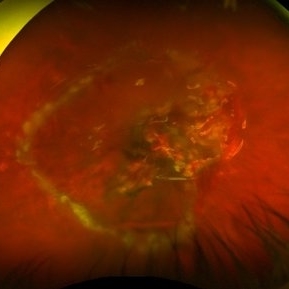
Advanced Diabetic Table-top TRD Intra-Operative Still undergoing Bimanual Vitrectomy Surgery
Photographer: Dr. Veer Singh
Imaging device: Intra-Operative Still
Condition/keywords: Diabetic Retinopathy, TRD, vitrectomy
-
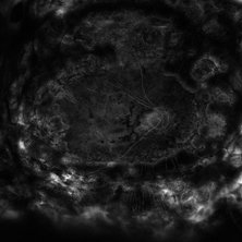
 Advanced Proliferative Diabetic Retinopathy
Apr 9 2025 by Gustavo Uriel Fonseca Aguirre
B-mode ultrasound of a patient with long-standing poorly controlled diabetes demonstrates characteristic findings of advanced proliferative diabetic retinopathy. The examination reveals moderate vitreous hemorrhage appearing as diffuse hyperechoic opacities throughout the vitreous cavity, along with a posterior hyaloid membrane densely infiltrated by hemorrhagic material, showing irregular thickening and increased reflectivity. A mild subhyaloid hemorrhage is visible as a subtle hyphema-like space anterior to the retinal surface. The study documents a total tractional retinal detachment, evidenced by rigid retinal folds with clear insertion points of vitreous strands, accompanied by a significant subretinal hemorrhage seen as a prominent hyperechoic collection beneath the elevated retina. These findings collectively illustrate the severe vitreoretinal interface pathology characteristic of end-stage diabetic eye disease, with predominant tractional components and distinct echographic stratification of hemorrhagic layers - from anterior vitreous involvement to deeper subretinal blood accumulation.
Photographer: Gustavo U. Fonseca Aguirre, Hospital Conde de Valenciana, Ciudad de México
Condition/keywords: diabetic retinopathy, tractional retinal detachment, Vitreous hemorrhage
-
 Bilateral CRVO and PDR
Nov 4 2021 by Stefanie Palmer
Patient with both PDR and CRVO, 34 year old female, post-COVID.
Photographer: Stefanie Palmer, CRA
Imaging device: Topcon
Condition/keywords: central retinal vein occlusion (CRVO), COVID-19, diabetic retinopathy, proliferative diabetic retinopathy (PDR), venous beading
-
 Bilateral Dengue Retinitis
Apr 5 2018 by JYOTI PATIL, Ph.D.
Right eye of a 40-year-old lady recovering from bilateral dengue retinitis shows dengue foveolitis, neovascularisation disc (NVD) and vitreous hemorrhage.
Photographer: Dr.Aditya Kelkar
Condition/keywords: bilateral dengue retinitis, diabetic retinopathy, vitreous hemorrhage
-
 Boat-Shaped Hemorrhage
Mar 1 2014 by Homayoun Tabandeh, MD, FASRS
Boat-shaped hemorrhage in a patient with retro-hyaloid hemorrhage associated with proliferative diabetic retinopathy.
Condition/keywords: diabetic retinopathy
-
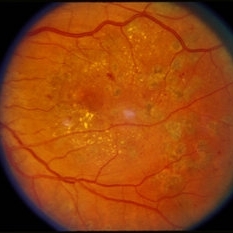 Color Photo of CSDME
Feb 19 2015 by H. Michael Lambert, MD
Color photo of CSDME after FALP.
Condition/keywords: clinically significant macular edema (CSME), color photo, diabetic retinopathy, FALP, laser
-
 Color Photo of CSDME
Feb 19 2015 by H. Michael Lambert, MD
Color photo of CSDME.
Condition/keywords: clinically significant macular edema (CSME), color photo, diabetic retinopathy, FALP
-
 Combined Tractional and Rhegmatogenous Retinal Detachment
Jan 30 2023 by Olivia Rainey
Ultra-widefield fluorescein angiography of a combined tractional and rhegmatogenous retinal detachment repair affecting the left eye. The retina is attached following silicone oil placement during most recent surgery. The patient was seeing CF at the time the image was taken.
Photographer: Olivia Rainey, OCT-C, COA
Imaging device: Optos California
Condition/keywords: diabetes, diabetic macular edema, diabetic retinopathy, fluorescein angiogram (FA), hyperfluorescence, right eye, scleral buckle, silicone oil, tractional retinal detachment, ultra-wide field imaging, ultra-widefield image
-
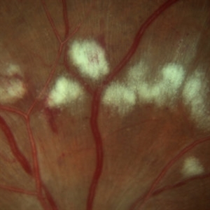
 Cotton Wool Spots
Mar 1 2014 by Homayoun Tabandeh, MD, FASRS
Cotton wool spots in a patient with hypertension and diabetes
Condition/keywords: cotton wool spots, diabetic retinopathy, hypertensive retinopathy
-
 Cotton Wool Spots
Mar 1 2014 by Homayoun Tabandeh, MD, FASRS
Cotton wool spots in a patient with hypertension and diabetes.
Condition/keywords: cotton wool spots, diabetic retinopathy, hypertensive retinopathy
-
Cutter based dissection in PDR
Oct 24 2022 by Manish Nagpal, MD, FRCS (UK), FASRS
This video highlights the ability of cutters to clear up the hemorrhage and remove attachments of proliferations flush on the retinal surface.
Photographer: Manish Nagpal
Condition/keywords: cutter, diabetic retinopathy, PDR, video, vitrectomy
-
 Cutter Segmentation in a case of Diabetic Combined Retinal Detachment | Intra-Operative Still
Apr 25 2023 by Veer Singh, MS, FVRS, FMRF, FICO (Retina)
Cutter Segmentation in a case of Diabetic Combined Retinal Detachment | Intra-Operative Still Patient underwent Vitrectomy with Silicone Oil
Photographer: Dr. Veer Singh
Condition/keywords: combined retinal detachment, cutter, diabetic retinopathy, intraoperative, pars plana vitrectomy (PPV)
-
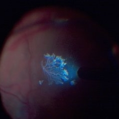 Detached NVE During PVD induction
Apr 27 2018 by Michael J. Koss, MD, PhD, MBA
A 73-year-old woman with macular pucker underwent a pars plana vitrectomy with membrane peeling. Additionally the patient suffers from diabetic retinopathy after being diagnosed with type 2 diabetes mellitus sixteen years ago. Prior to the procedure she was treated with a series of intravitreal Bevacizumab-injections due to diabetic macular edema. There was no history of a proliferative DRP. During the vitrectomy a branch of an obliterated NVE spontaneously detached and floated freely in the vitreous. The 3D shot was captured via Alcon’s NGENUITY® 3D Visualization System in form of photograph and video providing an outstandingly detailed image of the branched NVE.
Photographer: Michael Koss, Augenzentrum Nymphenburger Hoefe
Imaging device: Alcon’s NGENUITY® 3D Visualization System
Condition/keywords: diabetes, diabetic retinopathy, neovascularization elsewhere (NVE), pars plana vitrectomy (PPV), PVD induction
-
 Diabetic Macular Edema
Feb 12 2020 by DIEGO TOLENTINO
Proliferative diabetic retinopathy plus diabetic macular edema (cystoid).
Photographer: Diego Tolentino, CEOP
Condition/keywords: diabetic macular edema, diabetic retinopathy
-
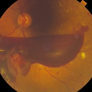
 Diabetic Macular TRD
Jan 10 2020 by Somnath Chakraborty, MD
Fundus Montage image of the left eye of a 48-year-old type 2 diabetic with post PRP macular extensive tractional retinal detachment involving macula.
Photographer: Pulak Roy
Condition/keywords: diabetic retinopathy, proliferative diabetic retinopathy (PDR), tractional retinal detachment, vitrectomy, vitreomacular surgery
-
 Diabetic Proliferative Retinopathy
Dec 1 2019 by Lucas Zago Ribeiro, MD
Fundus photograph of 75-year-old man with diabetic proliferative retinopathy with fibrovascular proliferation over the optic disc.
Photographer: Lucas Zago Ribeiro, Federal University of São Paulo
Imaging device: Zeiss Visucam 524
Condition/keywords: diabetic retinopathy, fibrovascular proliferation, neovascularization (NV)
-
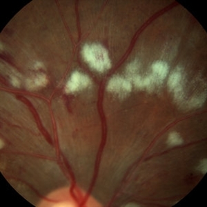
 Diabetic Retinopathy
Apr 5 2018 by JYOTI PATIL, Ph.D.
Fundus photograph of a 45-year-old man with diabetic retinopathy having severe cotton wool spot.
Photographer: Dr.Aditya Kelkar
Condition/keywords: diabetic retinopathy
-
 Diabetic Retinopathy
Dec 11 2019 by Lauren Whaley
44-year-old male diabetic patient had an acute change in A1C over 9 months and ended up with a tractional retinal detachmen in right eye. This photo is 2 weeks post operative with current vision level at hand motion. He had extensive laser, retinectomy, and silicone oil fill.
Photographer: Lauren R. Whaley, COA
Imaging device: Optos Wide Field
Condition/keywords: diabetes, diabetic retinopathy, fibrosis, laser scarring, proliferative vitreoretinopathy (PVR), retinectomy, silicone oil, tractional retinal detachment
-
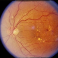
 Diabetic Retinopathy
Mar 14 2021 by Marco Antonio Sauza
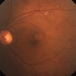
DR in a 40-year-old male with DM1.
Photographer: Marco Sauza
Imaging device: Zeiss
Condition/keywords: background diabetic retinopathy (BDR)
-
 Diabetic Retinopathy
Apr 5 2018 by JYOTI PATIL, Ph.D.
Fundus photograph with diabetic retinopathy observed with hemorrhage.
Photographer: Dr.Aditya Kelkar
Condition/keywords: diabetic retinopathy
-
 Diabetic Retinopathy
Apr 5 2018 by JYOTI PATIL, Ph.D.
Boat-shaped hemorrhage in a patient with retro-hyaloid hemorrhage associated with proliferative diabetic retinopathy.
Photographer: Dr.Aditya Kelkar
Condition/keywords: diabetic retinopathy
 Loading…