Search results (121 results)
-
 Abundant Hard Exudates - Diabetic Macular Edema
Oct 3 2013 by Gerardo Garcia-Aguirre, MD
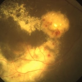
Abundant hard exudates - diabetic macular edema.
Condition/keywords: diabetic macular edema
-
 Angiographic Diabetic Macular Edema in a Case of Proliferative Diabetic Retinopathy
Apr 9 2024 by Akansha Sharma
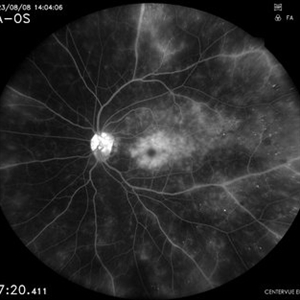
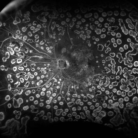
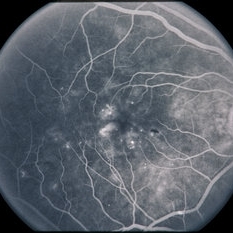
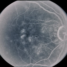
Fundus fluorescein angiographic image of 62 year old male demonstrating angiographic diabetic macular edema in a case of proliferative diabetic retinopathy.
Photographer: Dr. Akansha Sharma, Bharati Eye Hospital
Condition/keywords: clinically significant macular edema (CSME), diabetic blindness, diabetic macular edema, proliferative diabetic retinopathy (PDR)
-
 CIRCINATE RETINOPATHY
Oct 19 2022 by Akansha Sharma
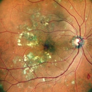
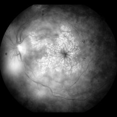
COLOUR FUNDUS PHOTOGRAPH OF A 51 YEAR OLD MALE WITH DIABETIC MACULOPATHY
Photographer: Dr. Akansha Sharma-Retina Foundation, Ahmedabad
Condition/keywords: circinate retinopathy, diabetic macular edema, diabetic maculopathy
-
 Clinically Significant Macular Edema
Apr 23 2015 by Mehul A Shah
Patient presented with complaints of diminished vision ou.
Photographer: Mehul Shah
Imaging device: Zeiss FF450 Plus
Condition/keywords: diabetic macular edema
-
 CME DME After CE
Aug 27 2014 by Susanna S. Park, MD, PhD
Macular OCT of a 62-year-old diabetic woman with severe vision loss 2 weeks after cataract surgery due to severe worsening of macular edema. Exam also showed new proliferative diabetic retinopathy.
Photographer: chandra
Condition/keywords: diabetic macular edema, optical coherence tomography (OCT)
-
 Combined Tractional and Rhegmatogenous Retinal Detachment
Jan 30 2023 by Olivia Rainey
Ultra-widefield fluorescein angiography of a combined tractional and rhegmatogenous retinal detachment repair affecting the left eye. The retina is attached following silicone oil placement during most recent surgery. The patient was seeing CF at the time the image was taken.
Photographer: Olivia Rainey, OCT-C, COA
Imaging device: Optos California
Condition/keywords: diabetes, diabetic macular edema, diabetic retinopathy, hyperfluorescence, right eye, scleral buckle, silicone oil, tractional retinal detachment, ultra-wide field imaging, ultra-widefield image
-
 Diabetic Macular Edema
May 28 2016 by Olivia Rainey
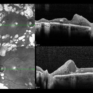
Optical coherence tomography of an 54-year-old female with diabetic macular edema affecting both eyes. Patient has a history of proliferative diabetic retinopathy s/p PRP/PPV/MP/EL, and glaucoma s/p tube shunt in both eyes. There has been a persistence of her macular edema and limited response to antiVEGF therapy, which puts into question whether there is another cause for her edema. Leading the possible causes is her renal insufficiency and fluid retention. Patient was seeing 20/50 in the right eye and 20/80 in the left eye.
Photographer: Olivia Rainey
Imaging device: Heidelberg Spectralis
Condition/keywords: anti-VEGF, diabetic macular edema, edema, glaucoma, optical coherence tomography (OCT), pan-retinal photocoagulation (PRP), proliferative diabetic retinopathy (PDR)
-
 Diabetic Macular Edema
Mar 14 2021 by Marco Antonio Sauza
Diabetic Macular Edema
Photographer: Marco Sauza
Imaging device: Heidelberg
Condition/keywords: diabetic macular edema
-
 Diabetic Macular Edema
Feb 12 2020 by DIEGO TOLENTINO
Proliferative diabetic retinopathy plus diabetic macular edema (cystoid).
Photographer: Diego Tolentino, CEOP
Condition/keywords: diabetic macular edema, diabetic retinopathy
-
 Diabetic Macular Edema
Feb 7 2024 by Virginia Gebhart
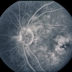
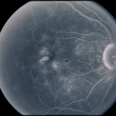
FA of 70 year old male with diabetic macular edema. FA shows early hyper-fluorescence with late leakage and capillary dropout in the temporal macula. Focal laser performed.
Photographer: Virginia Gebhart
Imaging device: Optos California
Condition/keywords: capillary dropouts, macular edema
-
 Diabetic Macular Edema
Apr 28 2025 by Gustavo Uriel Fonseca Aguirre
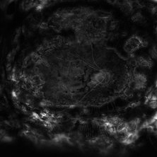
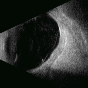
This B-mode longitudinal ultrasound scan demonstrates irregular macular thickening with homogeneous medium-to-high internal reflectivity, consistent with diabetic macular edema. The lesion shows poorly defined borders and absence of cystic spaces or subretinal fluid on dynamic evaluation.
Photographer: Gustavo U. Fonseca Aguirre, Hospital Conde de Valenciana, Ciudad de México
Condition/keywords: diabetic macular edema
-
 Diabetic Macular Edema
Jul 3 2025 by Gustavo Uriel Fonseca Aguirre
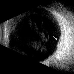
This B-mode longitudinal ultrasound scan demonstrates diabetic macular edema with mild subretinal fluid accumulation, appearing as a subtle hypoechoic space beneath the neurosensory retina. The macular region shows retinal thickening and heterogeneous medium reflectivity, consistent with active exudative changes (arrow). No vitreomacular traction is observed.
Photographer: Gustavo U. Fonseca Aguirre, Hospital Conde de Valenciana, Ciudad de México
Condition/keywords: diabetic macular edema
-
 Diabetic Macular Edema
Feb 12 2025 by Kimberly Wakester
Horizontal OCT scan of a 63-year-old woman with diabetic macular edema in the right eye. When reviewing the scan, one of the intraretinal cyst (IRC) appears heart shaped. A fun scan to see just a few day's before Valentine's day.
Photographer: Kimberly Wakester, COA
Imaging device: Heidelberg
Condition/keywords: diabetic macular edema, intraretinal cyst
-
 Diabetic Macular Edema
Oct 12 2012 by Gregg T. Kokame, MD, MMM, FASRS
Diabetic macular edema
Photographer: Jaclyn Pisano, Retina Consultants of Hawaii
Imaging device: Zeiss FF-450 plus
Condition/keywords: diabetic macular edema
-
---thumb.JPG/image-square;max$300,300.ImageHandler) Diabetic Macular Edema
Diabetic Macular Edema
Oct 26 2012 by Mallika Goyal, MD
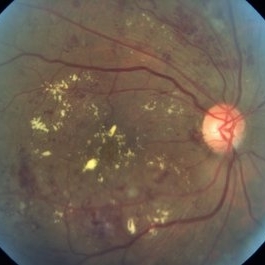
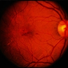
Fundus photograph of left eye of 55-year-old diabetic and hypertensive gentleman with normal serum lipids showing abundant foveal hard exudates.
Condition/keywords: diabetic macular edema
-
---thumb.JPG/image-square;max$300,300.ImageHandler) diabetic macular edema
diabetic macular edema
Oct 26 2012 by Mallika Goyal, MD
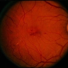
Fundus photograph of left eye of 58-year-old diabetic gentleman with normal serum lipids showing foveal hard exudates.
Condition/keywords: foveal hard exudates
-
 Diabetic Macular Edema
Feb 7 2014 by David Callanan, MD
56-year-old patient with diabetic macular edema.
Condition/keywords: diabetic macular edema
-
 Diabetic Macular Edema
Feb 7 2014 by David Callanan, MD
56-year-old patient with diabetic macular edema.
Condition/keywords: diabetic macular edema
-
 Diabetic Macular Edema
Feb 7 2014 by David Callanan, MD
56-year-old patient with diabetic macular edema.
Condition/keywords: diabetic macular edema
-
 Diabetic Macular Edema
Feb 7 2014 by David Callanan, MD
56-year-old patient with diabetic macular edema.
Condition/keywords: diabetic macular edema
-
 Diabetic Macular Edema
Feb 7 2014 by David Callanan, MD
56-year-old patient with diabetic macular edema.
Condition/keywords: diabetic macular edema
-
 Diabetic Macular Edema
Feb 7 2014 by David Callanan, MD
56-year-old patient with diabetic macular edema.
Condition/keywords: diabetic macular edema
-
 Diabetic Macular Edema
Feb 7 2014 by David Callanan, MD
56-year-old patient with diabetic macular edema.
Condition/keywords: diabetic macular edema
-
 Diabetic Macular Edema
Feb 7 2014 by David Callanan, MD
56-year-old patient with diabetic macular edema.
Condition/keywords: diabetic macular edema
-
 Diabetic Macular Edema
Feb 7 2014 by David Callanan, MD
56-year-old patient with diabetic macular edema.
Condition/keywords: diabetic macular edema
 Loading…