Search results (263 results)
-
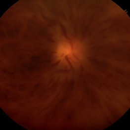
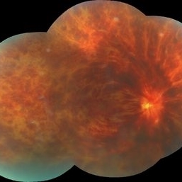
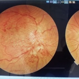
 Bilateral CRVO and PDR
Nov 4 2021 by Stefanie Palmer
Patient with both PDR and CRVO, 34 year old female, post-COVID.
Photographer: Stefanie Palmer, CRA
Imaging device: Topcon
Condition/keywords: central retinal vein occlusion (CRVO), COVID-19, diabetic retinopathy, proliferative diabetic retinopathy (PDR), venous beading
-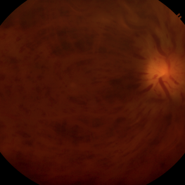
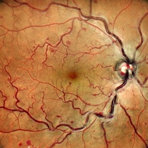 Blocked and Blurry: A Vein's Midlife Crisis
Jun 2 2025 by rohan jain
A color photograph showing central retinal vein occlusion
Photographer: Dr. ROHAN JAIN
Imaging device: mirante
Condition/keywords: central retinal vein occlusion (CRVO), crvo
-
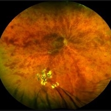
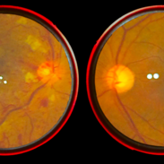
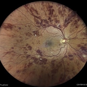
 Branch Retinal Artery Occlusion With Central Retinal Vein Occlusion
Jun 19 2019 by Unnati Vishwanath Shukla, M. S. ,DNB, FVRS FNERF, MNAMS,PhD Scholar(Retina)
A 65-year-old male hypertensive patient having resolving nonischemic central retinal vein occlusion presenting with inferotemporal branch retinal artery occlusion.
Photographer: Unnati Shukla
Condition/keywords: branch retinal artery occlusion (BRAO), central retinal vein occlusion (CRVO), cotton wool exudates, venous stasis
-
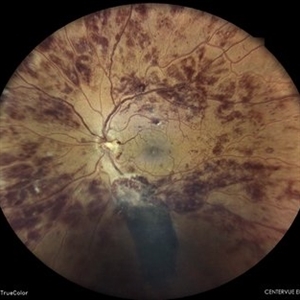
 Branch Retinal Artery Occlusion With Concurrent Central Retinal Vein Occlusion
Oct 5 2016 by Larry M Puthenparambil, MD
Branch retinal artery occlusion with concurrent central retinal vein occlusion.
Photographer: Stacey Groom
Imaging device: Topcon
Condition/keywords: branch retinal artery occlusion (BRAO), central retinal vein occlusion (CRVO)
-
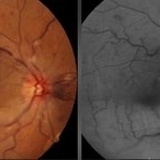
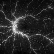
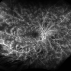
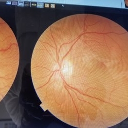
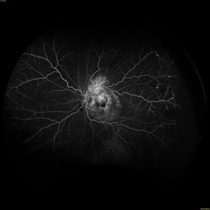
 Central Retinal Vein Occlusion
Jul 13 2018 by Olivia Rainey
Ultra-wide field fluorescein angiogram of a patient presenting with a central retinal vein occlusion their right eye.
Photographer: Olivia Rainey
Imaging device: Optos
Condition/keywords: central retinal vein occlusion (CRVO), fluorescein angiogram (FA), fluorescein leakage, hemorrhage, Optos, ultra-wide field imaging
-
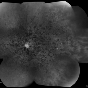
 Central Retinal Vein Occlusion
Jul 13 2018 by Olivia Rainey
Ultra-wide field, pseudocolor montage of a patient presenting with a central retinal vein occlusion, as well as, an inferior chorioretinal scar in their right eye.
Photographer: Olivia Rainey
Imaging device: Optos
Condition/keywords: central retinal vein occlusion (CRVO), chorioretinal scar, montage, Optos, pseudocolor, ultra-wide field imaging
-
 Central Retinal Vein Occlusion
Jan 21 2022 by Olivia Rainey
Ultra-widefield fluorescein angiogram of a 23-year-old female with a Central Retinal Vein Occlusion affecting her left eye. The patient presented on 12/22/2021 cc20/40-2 vision in the left eye. The patient reported recent trauma of being hit with a fist on both sides of face followed by vision loss. The patient has history of Hashimoto's thyroid disease. The following labs have been ordered, PT, PTT, CBC, antithrombin III activity, protein C, protein S, Factor V Leiden mutation, Prothrombin (G20210A), lipid panel, HbA1c, quantiferon gold, RPR, and CXR.
Photographer: Olivia Rainey, OCT-C, COA
Imaging device: Optos California
Condition/keywords: central retinal vein occlusion (CRVO), disc leakage, fluorescein angiogram (FA), fluorescein leakage, left eye, non-ischemic central retinal vein occlusion (CRVO), Optos, trauma, ultra-wide field imaging
-
 Central Retinal Vein Occlusion
Jan 26 2020 by Prithvi Chandrakanth
Transient visual obscuration in a 52-year-old male patient in his right eye, fundus examination revealed dot, blot and flame shaped hemorrhage with cotton wool patches.
Photographer: Dr.PRITHVI CHANDRAKANTH, ARAVIND EYE HOSPITAL, UDUMALPET
Imaging device: TRASH TO TREASURE RETCAM
Condition/keywords: central retinal vein occlusion (CRVO), retcam, smartphone fundus photography
-
 Central Retinal Vein Occlusion
Sep 14 2017 by Nichole Lewis
Central retinal vein occlusion.
Photographer: Nichole Lewis
Condition/keywords: central retinal vein occlusion (CRVO)
-
 Central Retinal Vein Occlusion
Jan 28 2024 by Gayathri Mohan
Fundus photograph of a 50 year old woman showing a CRVO.
Photographer: Dr Gayathri Mohan, Thumbay Medical and Dental Speciality Centre, U.A.E
Imaging device: 3nethra
Condition/keywords: central retinal vein occlusion (CRVO), ischemic CRVO
-
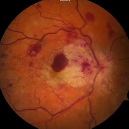
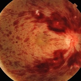
 Central Retinal Vein Occlusion
Nov 26 2020 by Priya Rasipuram Chandrasekaran, MBBS, DO, DNB, FRCS
A 44-year-old male patient presented with no underlying systemic illness presented with this picture showing extensive scattered superficial and deep retinal hemorrhages to confluent retinal hemorrhages extending to all the quadrants associated with marked dilatation and tortuosity of vessels and associated with optic disc edema, macular edema and retinal thickening giving the appearance of blood and thunder retina.
Condition/keywords: central retinal vein occlusion (CRVO)
-
 Central Retinal Vein Occlusion
Feb 20 2013 by From the Collections of Thomas M. Aaberg, MD and Thomas M. Aaberg Jr., MD
Retinal hemorrhages Central Retinal Vein Occlusion Fundus Photograph
Condition/keywords: central retinal vein occlusion (CRVO), retinal hemorrhage
-
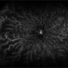
 Central Retinal Vein Occlusion
Feb 20 2021 by Stephanie Burke
Ultra wide-field FA image of a 22-year-old male with CRVO.
Photographer: Stephanie Burke, CRA ,OCT-C
Condition/keywords: central retinal vein occlusion (CRVO)
-
 Central Retinal Vein Occlusion
Nov 26 2020 by Priya Rasipuram Chandrasekaran, MBBS, DO, DNB, FRCS
A 44-year-old male patient presented with no underlying systemic illness presented with this picture showing extensive scattered superficial and deep retinal hemorrhages to confluent retinal hemorrhages extending to all the quadrants associated with marked dilatation and tortuosity of vessels and associated with optic disc edema, macular edema and retinal thickening giving the appearance of blood and thunder retina.
Condition/keywords: central retinal vein occlusion (CRVO)
-
 Central Retinal Vein Occlusion
Apr 9 2024 by Akansha Sharma
Color fundus photograph of a 73 year old hypertensive male with central retinal vein occlusion.
Photographer: Dr. Akansha Sharma, Bharati Eye Hospital
Condition/keywords: central retinal vein occlusion (CRVO), ischemic CRVO
-
 Central Retinal Vein Occlusion
Apr 9 2024 by Akansha Sharma
Color fundus photograph of a 73 year old hypertensive male with central retinal vein occlusion.
Photographer: Dr. Akansha Sharma, Bharati Eye Hospital
Condition/keywords: central retinal vein occlusion (CRVO), ischemic CRVO
-
 Central Retinal Vein Occlusion
Feb 22 2021 by AGNES KIM
Central Retina Vein Occlusion
Imaging device: Imagenet
Condition/keywords: central retinal vein occlusion (CRVO)
-
 Central Retinal Vein Occlusion
Feb 28 2021 by AGNES KIM
Fundus photograph of unilateral CRVO.
Photographer: Agnes Kim
Condition/keywords: central retinal vein occlusion (CRVO)
-
 Central Retinal Vein Occlusion
Sep 27 2024 by Jeffrey Barker
65 year old male with a Central Retinal Vein Occlusion and Macular Edema and Capillary Nonperfusion.
Photographer: Jeffrey P. Barker
Condition/keywords: central retinal vein occlusion (CRVO), macular edema
-
 Central Retinal Vein Occlusion
Jul 21 2024 by César Adrián Gómez Valdivia, MD
Central Retinal Vein Occlusion found in a 72 year old patient with history of uncontrolled Hypertension. Non-Ischemic Variant.
Photographer: Erika Paulina Ornelas Cazares
Imaging device: TOPCON TRC-50DX
Condition/keywords: central retinal vein occlusion (CRVO)
-
 Central Retinal Vein Occlusion
Sep 27 2024 by Korey Starkey
Fluorescein angiogram of a 75 year old patient with central retinal vein occlusion. FA shows areas of patchy ischemia and petaloid leakage. Patient is being treated with anti-vegf treatments at this time.
Photographer: Korey Starkey
Condition/keywords: central retinal vein occlusion (CRVO), FLUORESCEIN ANGIOGRAPHY, ischemia, macular edema, petaloid leakage, ultra-widefield image
-
 Central Retinal Vein Occlusion
Apr 13 2023 by Virginia Gebhart
68-year-old male with Central Retinal Vein Occlusion with Macular Edema. Pt presented with VA of count fingers @ 5 ft. Pt was treated with Avastin
Photographer: Virginia Gebhart, Retina Consultants of Carolina
Imaging device: Topcon TRC 50DX
Condition/keywords: central retinal vein occlusion (CRVO)
-
---thumb.jpg/image-square;max$300,300.ImageHandler) Central Retinal Vein Occlusion
Central Retinal Vein Occlusion
Oct 30 2012 by Lihteh Wu, MD
35-year-old hypertensive man with an acute CRVO. Notice the peripapillary cotton wool spots, superficial flame shaped hemorrhages and deeper dot and blot hemorrhages in all 4 quadrants. This is the typical blood and thunder appearance of a CRVO.
Condition/keywords: central retinal vein occlusion (CRVO), cotton wool spots
 Loading…