Search results (164 results)
-
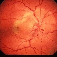
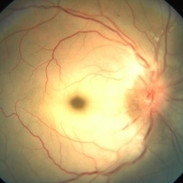
 24 Hours Post Scleral Wound Closure+ Scleral Buckle+25 g Vitrectomy+Silicon Oil
Jan 23 2015 by Carlos Quezada-Ruiz, MD, FASRS
24 hours post op fundus photograph of a 43-year-old man who had perforating injury to the right eye with a small piece of plastic while he was hammering. OD LP, subconjunctival hemorrhage, clear cornea, hyphema, irido and ciclodyalisis as well as a luxated lens with traumatic cataract and a dense vitreous hemorrhage. B-US showed rhegmatogenous retinal detachment with a tear and a big inferior hemorrhagic choroidal detachment. 360 peritomy revealed 2-entry scleral wounds were found in zone II (M V and M VI) and closure was performed. 25 G PPV was performed with the infusion canal placed in the AC through the limbus. Lensectomy and removal of a dense recent vitreous hemorrhage revealed a white detached retina with an exit wound through the temporal inferior segment of the optic nerve with a nasal GRT and sub retinal hemorrhage as well as temporal inferior choroidal, PVD was induced and PFOs helped stabilizing the retina while vitrectomy and sub-retinal hemorrhage was removed through the GRT. Fluid air exchange was made and 360 endolaser over the buckle indentation was done and silicon oil was used as endotamponade. This picture was taken 24 hrs after the surgery.
Photographer: Lilibeth Rodriguez, Instituto de la Visión. Torreon, Mexico.
Condition/keywords: central retinal artery occlusion (CRAO), giant retinal tear, trauma
-
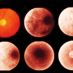
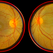
 Acute Central Retinal Artery Occlusion with Natural Reperfusion
Mar 12 2021 by Kushal S Delhiwala, MBBS, MS, FMRF,FICO, FAICO
Fundus photographs of 33-year-old healthy male with right eye acute CRAO of 12 hours duration showing cattle trucking, extensive retinal whitening and cherry red spot (left image). Right image 18 hours later showing reduced extent of retinal whitening and absent cattle trucking, suggestive of natural restoration of perfusion.
Photographer: Kushal Delhiwala, Netralaya superspeciality eye hospital, Ahmedabad, Gujarat,India
Imaging device: Optos Daytona
Condition/keywords: cattle trucking, central retinal artery occlusion (CRAO), cherry red spot
-
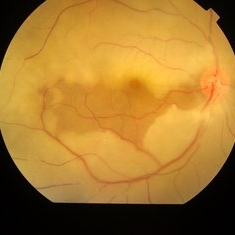
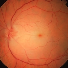
 AION With Ciliotretinal Artery Occlusion
May 2 2013 by Henry J. Kaplan, MD
AION accompanied by partial CRAO which is visible as retinal edema and cherry red spot.
Condition/keywords: anterior ischemic optic neuropathy, central retinal artery occlusion (CRAO)
-
 AO
AO
Aug 21 2013 by Howard Schatz, MD
CRAO.
Condition/keywords: AO, central retinal artery occlusion (CRAO)
-
 AO
AO
Aug 21 2013 by Howard Schatz, MD
Recent CRAO.
Condition/keywords: AO, central retinal artery occlusion (CRAO)
-
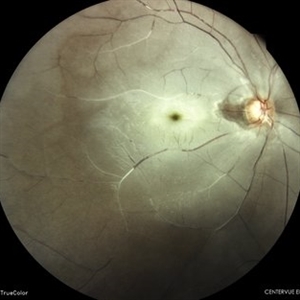
 Central Artery Occlusion
Aug 6 2023 by Anjana Mirajkar, MS Ophthalmology
Color photo of a 42 year old male in a case of central artery occlusion with cilio retinal artery sparing
Photographer: Dr. Anjana Mirajkar -Retina Foundation, Ahmedabad
Condition/keywords: Central Retinal Artery Occlusion, central retinal artery occlusion (CRAO)
-
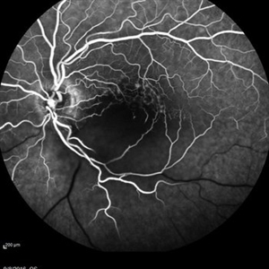
 Central Artery Occlusion with Cilio Retinal Artery Sparing
Aug 6 2023 by Anjana Mirajkar, MS Ophthalmology
Wide field view of FA (Late phase) of a 42 year old male in a case of central artery occlusion with cilio retinal artery sparing showing delayed arterial filling with choroidal filling
Photographer: Dr. Anjana Mirajkar -Retina Foundation, Ahmedabad
Condition/keywords: central retinal artery occlusion (CRAO)
-
 Central Artery Occlusion with Cilio Retinal Artery Sparing
Aug 6 2023 by Anjana Mirajkar, MS Ophthalmology
Central FA frame (Late phase) of a 42 year old male in a case of central artery occlusion with cilio retinal artery sparing
Photographer: Dr. Anjana Mirajkar -Retina Foundation, Ahmedabad
Condition/keywords: central retinal artery occlusion (CRAO)
-
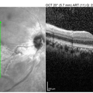 Central Artery Occlusion with Cilio Retinal Artery Sparing
Aug 6 2023 by Anjana Mirajkar, MS Ophthalmology
OCT image (Vertical scan ) of RE of a 42 year old male in a case of central artery occlusion with cilio retinal artery sparing showing loss of differentiation of retinal layers in nasal half.
Photographer: Dr. Anjana Mirajkar -Retina Foundation, Ahmedabad
Condition/keywords: central retinal artery occlusion (CRAO)
-
 Central retinal artery occlusion
Nov 30 2022 by Ethan K Sobol, MD
A central retinal artery occlusion with cilioretinal artery sparing, imaged using a Volk Panretinal 2.2 and an iPhone camera in the emergency department.
Photographer: Jared Raabe, MD, Emory University Hospital
Imaging device: IPhone 13 Pro
Condition/keywords: central retinal artery occlusion (CRAO)
-
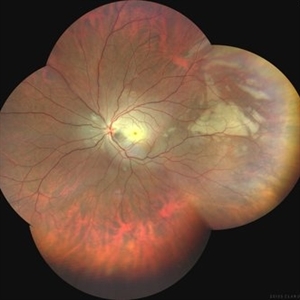 Central Retinal Artery Occlusion
Mar 21 2025 by T. P . VIGNESH, MBBS,MS
Fundus photo montage of a 29 year old man with Central retinal artery occlusion.
Photographer: Bharathi
Imaging device: Zeiss Clarus
Condition/keywords: central retinal artery occlusion (CRAO)
-
 Central Retinal Artery Occlusion
Mar 11 2024 by Dr.Pavithra Subramanian
A 51 year old male with defective vision in right eye for past 4days.On examination RE RAPD present and Fundus examination found to be Right eye Central retinal artery occlusion with Grade 4 Hypertensive Retinopathy.
Photographer: Dr Pavithra Subramanian
Condition/keywords: central retinal artery occlusion (CRAO), malignant hypertension
-
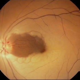
 Central Retinal Artery Occlusion
Apr 10 2024 by Tejaswita Verma
Left eye fundus photo of a 75 year old male with pale edematous retina with cherry red spot in a case of central retinal artery occlusion.
Photographer: DR. TEJASWITA VERMA
Imaging device: MIRANTE
Condition/keywords: central retinal artery occlusion (CRAO), cherry red spot
-
 Central Retinal Artery Occlusion
Mar 26 2019 by Gary R. Cook, MD, FACS
61-year-old male patient with acute CRAO OS demonstrating a hyperemic optic disc with a couple of peripapillary hemorrhages, generalized arteriolar narrowing, a cherry-red spot in the macula, and retinal whitening surrounding the fovea; VA= LP.
Imaging device: Topcon VT-50
Condition/keywords: central retinal artery occlusion (CRAO), cherry red spot, retinal whitening
-
 Central Retinal Artery Occlusion
Jan 22 2021 by Renata Garcia Franco, Md
65-year-old male, history of uncontrolled systemic arterial hypertension. Fluorescein angiography (FA) shows a delay in filling of the retinal arteries.
Photographer: Fatima Hernandez, Instituto de la Retina del Bajio SC
Imaging device: Zeiss
Condition/keywords: central retinal artery occlusion (CRAO)
-
 Central Retinal Artery Occlusion
Jan 22 2021 by Renata Garcia Franco, Md
65-year-old male, history of uncontrolled systemic arterial hypertension. Segmentation of blood in retinal arterioles, retinal whitening and cherry red spot.
Photographer: Fatima Hernandez, Instituto de la Retina del Bajio SC
Imaging device: Zeiss
Condition/keywords: central retinal artery occlusion (CRAO)
-
 Central Retinal Artery Occlusion
Feb 20 2013 by From the Collections of Thomas M. Aaberg, MD and Thomas M. Aaberg Jr., MD
No history; documented over time.
Condition/keywords: central retinal artery occlusion (CRAO)
-
 Central Retinal Artery Occlusion
Jun 4 2019 by Unnati Vishwanath Shukla, M. S. ,DNB, FVRS FNERF, MNAMS,PhD Scholar(Retina)
A young female patient of Indian origin on Oral Contraceptive medication presenting with Central Retinal Artery Occlusion with Cilioretinal artery Sparing.
Photographer: Unnati Shukla, C.H. Nagri Eye Hospital, NHL medical college, Ahmedabad,Gujarat,India.
Condition/keywords: central retinal artery occlusion (CRAO), cherry red spot, cilioretinal sparing, pale retina
-
 Central Retinal Artery Occlusion
Jan 13 2020 by Prithvi Chandrakanth
37-years-old male with complaints of sudden diminution of vision in the left eye for the past three days. Fundus examination revealed pale retina in the left eye with cherry red spot and normal fundus picture in right eye.
Photographer: DR.PRITHVI CHANDRAKANTH, ARAVIND EYE HOSPITAL, UDUMALPET
Imaging device: TRASH TO TREASURE RETCAM
Condition/keywords: central retinal artery occlusion (CRAO), cherry red spot, retcam, smartphone fundus photography
-
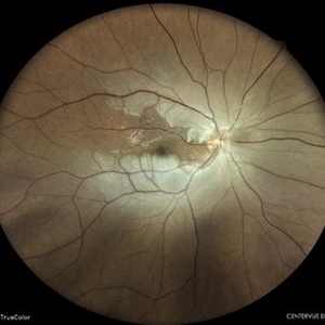 Central Retinal Artery Occlusion
May 7 2024 by Akansha Sharma
Color fundus photograph of a 40 year old female with central retinal artery occlusion.
Photographer: Dr. Akansha Sharma, Bharati Eye Hospital
Condition/keywords: central retinal artery occlusion (CRAO), CRAO
-
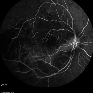
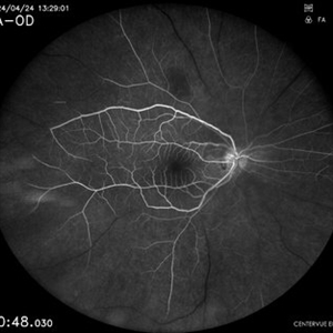 Central Retinal Artery Occlusion
May 7 2024 by Akansha Sharma
Early phase fluorescein angiography of a 40 year old female with central retinal artery occlusion.
Photographer: Dr. Akansha Sharma, Bharati Eye Hospital
Condition/keywords: central retinal artery occlusion (CRAO), CRAO
-
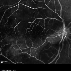
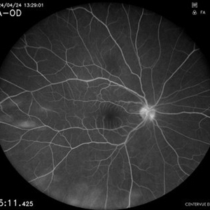 Central Retinal Artery Occlusion
May 7 2024 by Akansha Sharma
Late phase fluorescein angiography of a 40 year old female with central retinal artery occlusion.
Photographer: Dr. Akansha Sharma, Bharati Eye Hospital
Condition/keywords: central retinal artery occlusion (CRAO), CRAO
-
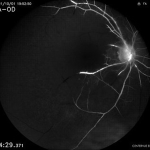 Central Retinal Artery Occlusion
Apr 17 2024 by Akansha Sharma
Fluorescein angiography of a 48 year old male with central retinal artery occlusion.
Photographer: Dr. Akansha Sharma, Bharati Eye Hospital
Condition/keywords: central retinal artery occlusion (CRAO), CRAO
-
 Central Retinal Artery Occlusion
Apr 17 2024 by Akansha Sharma
Color fundus photograph of a 48 year old male with central retinal artery occlusion.
Photographer: Dr. Akansha Sharma, Bharati Eye Hospital
Condition/keywords: central retinal artery occlusion (CRAO), CRAO
-
 Central Retinal Artery Occlusion
May 16 2017 by Olivia Rainey
Fluorescein angiogram of an 66-year-old female with a central retinal artery occlusion affecting her left eye.
Photographer: Olivia Rainey
Imaging device: Heidelberg Spectralis
Condition/keywords: 50 degrees, central retinal artery occlusion (CRAO), fluorescein angiogram (FA), left eye, mid phase, retinal ischemia
 Loading…