Search results (13 results)
-
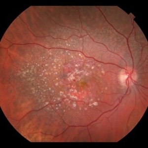 AMD With Calcified Drusen and Small, Deep IRH
Jul 22 2018 by John S. King, MD
AMD with calcified drusen and small, deep IRH
Photographer: Stacey
Imaging device: Topcon
Condition/keywords: calcified drusen
-
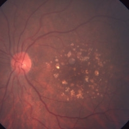
 Calcified Drusen
Mar 1 2014 by Homayoun Tabandeh, MD, FASRS
Calcified drusen in a patient with dry age-related macular degeneration.
Condition/keywords: age-related macular degeneration (AMD), calcified drusen
-
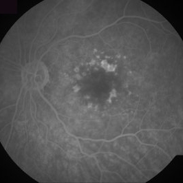 Calcified Drusen, Fluorescein Angiogram
Aug 23 2012 by Gerardo Garcia-Aguirre, MD
Fluorescein angiogram of a 72 year-old patient with dry age-related macular degeneration and calcified drusen.
Photographer: Noemí Hernández, Asociación para Evitar la Ceguera en México
Imaging device: Zeiss FF4
Condition/keywords: age-related macular degeneration (AMD), calcified drusen, dry age-related macular degeneration (dry AMD)
-
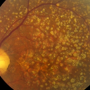
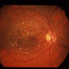
 Calcified drusen, fundus photograph
Aug 23 2012 by Gerardo Garcia-Aguirre, MD
Fundus photograph showing multiple white-yellowhish lesions corresponding to calcified drusen.
Photographer: Noemí Hernández, Asociación para Evitar la Ceguera en México
Imaging device: Zeiss FF4
Condition/keywords: calcified drusen, dry age-related macular degeneration (dry AMD)
-
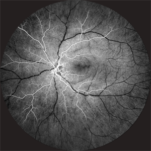
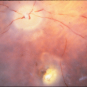
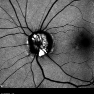 Drusen of Optic Disc
Mar 6 2018 by JEFFERSON R SOUSA, Tecg.º (Biomedical Systems Technology)
Female patient, 37 years old, Caucasian, with complaint of low lateral stroke in abos the eyes. In the retinal mapping examination and retinography, important alterations in the optic nerve head suggestive of Drusen PAPILLA were observed. After being confirmed in the Autofluorescence examination, we observed Autohyperfluorescence compatible with deposits of calcified hyaline material, as well as another complementary exam such as USG and OCT.
Photographer: JEFFERSON R SOUSA - Study Center and Ophthalmological Research Dr. Andre M V Gomes, Institute Dr. Suel Abujamra, Clinic Marco Antonio Albhy Ophthalmology, São Paulo-Brazil
Imaging device: Heidelberg - HRA Angiograph, Autofluorescence com 30 degrees.
Condition/keywords: calcified drusen, drusen of optic disc
-
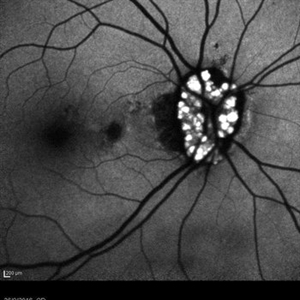 Drusen of Optic Disc
Mar 6 2018 by JEFFERSON R SOUSA, Tecg.º (Biomedical Systems Technology)
Female patient, 37 years old, Caucasian, with complaint of low lateral stroke in abos the eyes. In the retinal mapping examination and retinography, important alterations in the optic nerve head suggestive of DRUSAS DE PAPILA were observed. After being confirmed in the Autofluorescence examination, we observed Autohyperfluorescence compatible with deposits of calcified hyaline material, as well as another complementary exam such as USG and OCT.
Photographer: JEFFERSON R SOUSA - Study Center and Ophthalmological Research Dr. Andre M V Gomes, Institute Dr. Suel Abujamra, Clinic Marco Antonio Albhy Ophthalmology / São Paulo-Brazil
Imaging device: Heidelberg - HRA Angiograph, Autofluorescence com 30 degrees.
Condition/keywords: calcified drusen, drusen of optic disc
-
 Dry AMD
Jun 4 2014 by Henry J. Kaplan, MD
Multiple hard and calcified drusen. #1
Condition/keywords: age-related macular degeneration (AMD), calcified drusen, drusen, dry age-related macular degeneration (dry AMD)
-
 Slide 9-86
Feb 26 2019 by Lancaster Course in Ophthalmology
Calcified drusen.
Condition/keywords: calcified drusen
-
 Takayasu Retinopathy
Apr 30 2025 by Vishal Agrawal, MD, FRCS,FACS,FASRS
Fundus fluorescein angiography image of a young girl with diagnosed Takayasu arteritis who presented with complains of diminished vision in both eyes. FFA shows complete absence of venous filling with segmented blood column secondary to CRAO with peripheral avascular area.
Photographer: Dr Ayushi Gupta
Imaging device: Clarus 700
Condition/keywords: calcified drusen, CRAO, takayasu arteritis
-
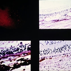
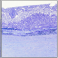
 Best Vitelliform Macular Dystrophy
Dec 10 2020 by McGill University Health Centre
Postmortem eyes from 101-year-old female. Past clinical history includes a poor vision for many years due to macular degeneration. The last visual acuity test recorded 6/15 OD and 6/6 OS. IOP 14 and 18 torr OS. Histopathology: Disclosed and yellow 2x2mm macular lesion. Microscopic examination: elevated placoid macular lesion with overlying pigment granules. Electron microscopy examination: pigment granules with abundant lipofuscin and melanolysosomes, photoreceptor cells markedly attenuated (less degenerated at the periphery) Numerous calcified drusen throughout the retina particularly in the posterior pole. RPE lipofuscin content is elevated in Best’s dystrophy. The extractability of the PRE lipofuscin fluorophores is reduced (it is normal during senescence). The defect in Best’s dystrophy accelerates this age related change in lipofuscin.
Condition/keywords: Best vitelliform macular dystrophy (BVMD), fundus photograph
-
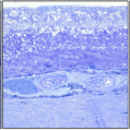
 Best Vitelliform Macular Dystrophy
Dec 10 2020 by McGill University Health Centre
Postmortem eyes from 101-year-old female. Past clinical history includes a poor vision for many years due to macular degeneration. The last Visual acuity test recorded 6/15 OD and 6/6 OS. IOP 14 and 18 torr OS. Histopathology: Disclosed and yellow 2x2mm macular lesion. Microscopic examination: elevated placoid macular lesion with overlying pigment granules. Electron microscopy examination: pigment granules with abundant lipofuscin and melanolysosomes, photoreceptor cells markedly attenuated (less degenerated at the periphery) Numerous calcified drusen throughout the retina particularly in the posterior pole. RPE lipofuscin content is elevated in Best’s dystrophy. The extractability of the PRE lipofuscin fluorophores is reduced (it is normal during senescence). The defect in Best’s dystrophy accelerates this age related change in lipofuscin.
Condition/keywords: Best vitelliform macular dystrophy (BVMD), histopathology, pathology
-
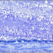
 Best Vitelliform Macular Dystrophy
Dec 10 2020 by McGill University Health Centre
Postmortem eyes from 101-year-old female. Past clinical history includes a poor vision for many years due to macular degeneration. The last visual acuity test recorded 6/15 OD and 6/6 OS. IOP 14 and 18 torr OS. Histopathology: Disclosed and yellow 2x2mm macular lesion. Microscopic examination: elevated placoid macular lesion with overlying pigment granules. Electron microscopy examination: pigment granules with abundant lipofuscin and melanolysosomes, photoreceptor cells markedly attenuated (less degenerated at the periphery) Numerous calcified drusen throughout the retina particularly in the posterior pole. RPE lipofuscin content is elevated in Best’s dystrophy. The extractability of the PRE lipofuscin fluorophores is reduced (it is normal during senescence). The defect in Best’s dystrophy accelerates this age related change in lipofuscin.
Condition/keywords: Best vitelliform macular dystrophy (BVMD), histopathology, pathology
-
 Best Vitelliform Macular Dystrophy
Dec 10 2020 by McGill University Health Centre
Postmortem eyes from 101-year-old female. Past clinical history includes a poor vision for many years due to macular degeneration. The last visual acuity test recorded 6/15 OD and 6/6 OS. IOP 14 and 18 torr OS. Histopathology: Disclosed and yellow 2x2mm macular lesion. Microscopic examination: elevated placoid macular lesion with overlying pigment granules. Electron microscopy examination: pigment granules with abundant lipofuscin and melanolysosomes, photoreceptor cells markedly attenuated (less degenerated at the periphery) Numerous calcified drusen throughout the retina particularly in the posterior pole. RPE lipofuscin content is elevated in Best’s dystrophy. The extractability of the PRE lipofuscin fluorophores is reduced (it is normal during senescence). The defect in Best’s dystrophy accelerates this age related change in lipofuscin.
Condition/keywords: Best vitelliform macular dystrophy (BVMD), histopathology, pathology
 Loading…