Search results (12 results)
-
---thumb.jpg/image-square;max$300,300.ImageHandler) Age Related Macular Degeneration
Age Related Macular Degeneration
May 3 2013 by Suber S. Huang, MD, MBA, FASRS
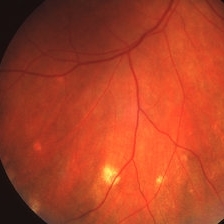
Age related macular degeneration.
Condition/keywords: advanced geographic atrophy, atrophic scar, atrophic spot, geographic atrophy, macula lesion, pigment epithelial atrophy, red-free, window defect
-
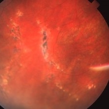
---thumb.jpg/image-square;max$300,300.ImageHandler) Age Related Macular Degeneration - Geographic Atrophy
Age Related Macular Degeneration - Geographic Atrophy
May 3 2013 by Suber S. Huang, MD, MBA, FASRS
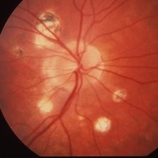
Geographic Atrophy.
Imaging device: Retina Diseases Imaging Analysis Reading Center
Condition/keywords: advanced geographic atrophy, atrophic scar, atrophic spot, geographic atrophy, macula lesion, pigment epithelial atrophy
-
---thumb.jpg/image-square;max$300,300.ImageHandler) Age Related Macular Degeneration - Geographic Atrophy
Age Related Macular Degeneration - Geographic Atrophy
May 3 2013 by Suber S. Huang, MD, MBA, FASRS
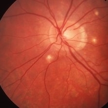
Geographic Atrophy.
Imaging device: Retina Diseases Imaging Reading Center
Condition/keywords: advanced geographic atrophy, atrophic scar, atrophic spot, geographic atrophy, macula lesion, pigment epithelial atrophy, red-free, window defect
-
---thumb.jpg/image-square;max$300,300.ImageHandler) Age Related Macular Degeneration - Geographic Atrophy
Age Related Macular Degeneration - Geographic Atrophy
May 3 2013 by Suber S. Huang, MD, MBA, FASRS
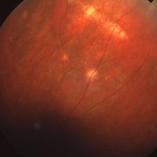
Geographic Atrophy.
Imaging device: Retina Diseases Imaging Analysis Reading Center
Condition/keywords: advanced geographic atrophy, atrophic scar, atrophic spot, geographic atrophy, macula lesion, pigment epithelial atrophy
-
 Atrophic Histoplasmosis Spots
Mar 27 2019 by Gary R. Cook, MD, FACS
Atrophic histo spots in the mid-periphery OD of an adult white male.
Imaging device: Topcon VT-50
Condition/keywords: atrophic spot, ocular histoplasmosis syndrome (OHS), presumed ocular histoplasmosis syndrome (POHS)
-
 Histoplasmosis
Mar 27 2019 by Gary R. Cook, MD, FACS
24-year-old white female with presumed ocular histoplasmosis (POHS) demonstrating some peripapillary atrophy and multiple atrophic histo spots around the optic nerve of her right eye; the patient was asymptomatic; V.A.= 20/20.
Imaging device: Topcon VT-50
Condition/keywords: atrophic spot, ocular histoplasmosis syndrome (OHS), peripapillary atrophy, presumed ocular histoplasmosis syndrome (POHS)
-
 Histoplasmosis
Mar 27 2019 by Gary R. Cook, MD, FACS
24-year-old white female with presumed ocular histoplasmosis (POHS) demonstrating minimal peripapillary atrophy but 3 atrophic histo spots around the optic nerve of her left eye; patient was asymptomatic; V.A.= 20/20.
Imaging device: Topcon VT-50
Condition/keywords: atrophic spot, ocular histoplasmosis syndrome (OHS), presumed ocular histoplasmosis syndrome (POHS)
-
 Linear Histoplasmosis
Mar 27 2019 by Gary R. Cook, MD, FACS
Middle-aged white male with presumed ocular histoplasmosis (POHS) demonstration single astrophic histo spots and a linear histo streak in the superior periphery.
Imaging device: Topcon VT-50
Condition/keywords: atrophic spot, ocular histoplasmosis syndrome (OHS), presumed ocular histoplasmosis syndrome (POHS)
-
 Linear Histoplasmosis Streaks
Mar 27 2019 by Gary R. Cook, MD, FACS
26-year-old white female with POHS showing multiple linear histo streaks in the periphery; V.A.= 20/200.
Imaging device: Topcon VT-50
Condition/keywords: atrophic, atrophic spot, ocular histoplasmosis syndrome (OHS), presumed ocular histoplasmosis syndrome (POHS)
-
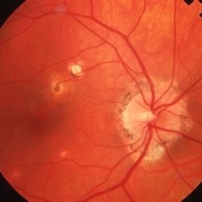 Ocular Histoplasmosis
Mar 27 2019 by Gary R. Cook, MD, FACS
Fellow eye (OD) of a 41-year-old white female with ocular histoplasmosis showing peripapillary atrophy and several atrophic histo spots OD; no CNVM present; V.A.= 20/20.
Imaging device: Topcon VT-50
Condition/keywords: atrophic spot, histoplasmosis, peripapillary atrophy, presumed ocular histoplasmosis syndrome (POHS)
-
 Scleral Indentation
Nov 9 2012 by Norman Byer
The next three photographs are of the same lesion in a 26-year-old man and demonstrate the value of scleral indentation. This view without indentation shows only a tiny pigmented and atrophic spot in the fundus.
Condition/keywords: atrophic spot, pigmented lesion, scleral indentation
-
 Scleral Indentation
Nov 9 2012 by Norman Byer
This is the same lesion as in the previous photograph. As scleral indentation begins, we see that the former tiny spot is actually the center of a larger whiter zone of uncertain etiology.
Condition/keywords: atrophic spot, scleral indentation, white retinal lesion
 Loading…