Search results (5 results)
-
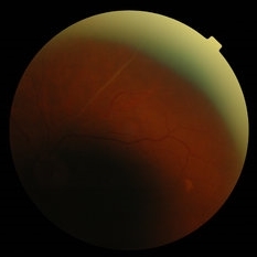
 AV Anastomosis
Sep 19 2017 by Purva Patwari
54-year-old female post cataract surgery.
Photographer: Dr Purva Patwari, Patwari Retina Center,Ahmedabad
Imaging device: Zeiss visu 500
Condition/keywords: arteriovenous anastomosis
-
 Branch Arterial Occlusion
Apr 12 2018 by Marco D'Angelo
Branch artery occlusion with collateral circulation (arterio-arterial vascular anastomosis) around the residual perfused branch. Left eye, 76-years-old male patient, normal visual acuity (20/20).
Photographer: Dr. Marco D'Angelo, S.Chiara Hospital, Trento, Italy
Imaging device: Topcon TRC-NW 6S
Condition/keywords: arteriovenous anastomosis, collateral retinal vessel, ghost vessels
-
 Retinal Arteriovenous Malformation
Jun 6 2020 by Albert Li, MD, FASRS
Montaged infrared retinal imaging of a 37-year-old asymptomatic man with a grade II arteriovenous malformation (AVM) in the nasal mid-periphery. The presentation of the AVM can be classified with three categories. Grade 1 AVMs are characterized by an abnormal capillary plexus between the major communicating vessels. Grade 2 AVMs are defined by the direct arteriovenous communication without the interposition of arterioles or capillaries. Grade 3 AVMs are characterized by widespread, large caliber anastomosing vessels that are associated with decreased visual acuity and intracranial AVMs. Because retinal AVMs are mostly asymptomatic and non-progressive, further testing may not be indicated unless there are concomitant neurological signs and symptoms or if there is a strong clinical suspicion of a grade 3 retinal AVM. Observation was recommended for the patient in this image. On his most recent follow-up at four months, the patient remained asymptomatic with a stable appearance of the lesion.
Imaging device: Heidelberg Spectralis
Condition/keywords: arteriovenous anastomosis, arteriovenous malformation
-
 Proliferative Diabetic Retinopathy with Temporal Seafan NVE Dragging Retinal Vein
Feb 15 2018 by Kushal S Delhiwala, MBBS, MS, FMRF,FICO, FAICO
58-year-old diabetic male presenting with Bilateral Proliferative diabetic retinopathy and centre involving Diabetic macular edema.Left eye fundus photograph showing large seafan NVE temporal to macula causing upward dragging of inferotemporal retinal vein and arteriovenous anastomosis.
Photographer: Dr Kushal Delhiwala, Netralaya superspeciality eye hospital, Ahmedabad
Imaging device: Zeiss Visucam 500
Condition/keywords: neovascularization elsewhere (NVE), proliferative diabetic retinopathy (PDR), sea fan
-
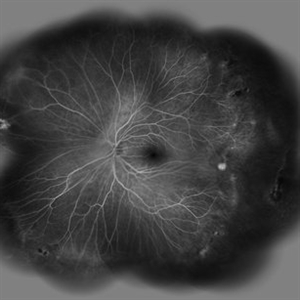 Vascular Abnormalities in an Asymptomatic Eye with FEVR
Jul 7 2015 by Hamid Ahmadieh, MD
Wide -field FA image of the asymptomatic left eye of a 28-year-old man with total RD secondary to FEVR in his right eye. Retinal vascular abnormalities including avascular zone in the extreme periphery associated with vasodilation and arteriovenous anastomosis are visible in the left eye.
Photographer: Soulmaz Shahmohammad, Negah Eye Center, Tehran, Iran
Imaging device: Specteralis
Condition/keywords: familial exudative vitreoretinopathy (FEVR), peripheral retinal nonperfusion
 Loading…