Search results (2962 results)
-
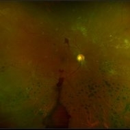
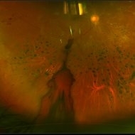
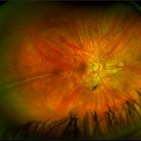
 Acute Central Retinal Artery Occlusion
Jul 27 2022 by Becca Harris
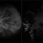
Ultra widefield FA/ICG of a 24 year old female with an acute central retinal artery occlusion affecting the right eye. Patient presented with extreme headaches following DAVF surgery the previous day. Patient has Factor VIII deficiency and had a cerebral venous thrombosis 9 years ago and lost vision in the right eye at that time. Patient has history of optic sheath fenestration OU and craniotomy. On initial evaluation, she had a CRAO as well as diffuse choroidal nonperfusion noted on optos FA. Suspect nonperfusion to third and sixth nerve leading to palsy. Occlusion of vasculature in the setting of recent endovascular embolization of fistulas in the CNS. Discussed diagnosis and poor prognosis with parents and patient. Patient had no light perception at the time of her initial appointment.
Photographer: Becca Harris
Imaging device: Optos California
Condition/keywords: Choroidal non-perfusion, fluorescein angiogram (FA), indocyanine green (ICG) angiography, non-perfusion, Optos, Right Eye, ultra-wide field imaging
-
 Central Serous Retinopathy
Mar 19 2024 by Corey Grant
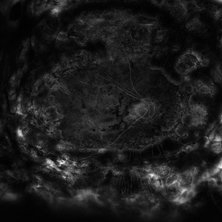
Ultra Wide-Field Fundus Autofluorescence Imaging of a 37 year old female with Central Serous Retinopathy affecting her right eye. Patient Visual Acuity was 20/20 in both eyes. Patient reported black spots in her vision onset three years ago, with associating flashes of light. Patient reports history of cortisone back injections a few years ago and denies Flonase use. The physician stated that there is hyperautofluorescence in the area of gutter of Sub-Retinal Fluid which likely happened from CSR.
Photographer: Corey Grant, OSC
Imaging device: OPTOS CALIFORNIA RGB
Condition/keywords: Central Serous Chorioretinopathy (CSR), central serous retinopathy (CSR), fundus autofluorescence (FAF), Guttering, hyperautofluorescence, inferior retina, OPTOS, Retina, Right Eye, subretinal fluid, ULTRA WIDE FIELD
-
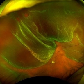
 Combined Tractional and Rhegmatogenous Retinal Detachment
Jan 30 2023 by Olivia Rainey
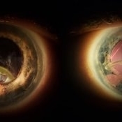
Ultra-widefield fluorescein angiography of a combined tractional and rhegmatogenous retinal detachment repair affecting the left eye. The retina is attached following silicone oil placement during most recent surgery. The patient was seeing CF at the time the image was taken.
Photographer: Olivia Rainey, OCT-C, COA
Imaging device: Optos California
Condition/keywords: diabetes, diabetic macular edema, diabetic retinopathy, fluorescein angiogram (FA), hyperfluorescence, right eye, scleral buckle, silicone oil, tractional retinal detachment, ultra-wide field imaging, ultra-widefield image
-
 Congenital Nuclear Cataract
Jul 5 2024 by Zach Seim
This is a slit-lamp photograph of a 10 year old female with a congenital nuclear cataract OD. Patient presented with VA Dsc 20/200. Patient was counseled on surgical options.
Photographer: Zach Seim
Imaging device: Slit Lamp Photography on Samsung Galaxy 7
Condition/keywords: cataract, congenital cataract, nuclear sclerosis, right eye, slit lamp photo
-
 Dislocated Crystalline Lens
Mar 19 2024 by Annaka Gooding
Ultra Wide field fundus photography of a 70 year old male who presented to clinic with a sudden increase of vision due to dropped crystalline lens secondary to severely dense cataract. Patient reported seeing a full black circle in his inferior visual field. Patient's visual acuity at time of visit was 20/100 with a +5.00 diopter lens. The physician recommended surgical intervention, and discussed surgery for PPV/PPL/IOL implantation with an ACIOL.
Photographer: Annaka Gooding, CPO
Imaging device: Optos California RGB
Condition/keywords: dislocated crystalline lens, fundus photography, inferior retina, OPTOS CALIFORNIA RGB, Right Eye, Ultra-wide field retinal imaging
-
 Dislocated IOL
Jun 4 2024 by Marlee Curnutt
Slit lamp photo of a 64 year old woman presenting with worsening vision and depth perception after trauma induced by a dog, which dislocated her IOL. The patient's IOL haptic was seen in the AC, and almost abutting cornea. Patient's vision upon presentation was DCC CF@1 feet. Patient was counseled and underwent an IOL exchange.
Photographer: Marlee Curnutt, COA
Imaging device: Galaxy A42
Condition/keywords: dislocated intraocular lens (IOL), haptic, IOL, right eye, slit lamp photo, slit lamp photography, trauma
-
 Idiopathic Choroidal Neovascularization
Mar 2 2023 by Corey Grant
Optical coherence tomography and ultra-wide field fundus photograph of a 51 year old male with idiopathic choroidal neovascularization affecting his right eye. The patient had no symptoms at the time of the appointment and his vision was Dcc20/20-2 OU. The physcian stated that there wasn't active exudation on the exam or ocular imaging and based on the clinical findings, he has recommended we defer any treatments.
Photographer: Corey Grant
Imaging device: Heidelberg Spectralis, OPTOS California
Condition/keywords: choroidal neovascularization (CNV), CNVM, fundus photograph, OCT, optical coherence tomography (OCT), Optos, Right Eye, ultra-wide field imaging
-
 Iris Nevus
Jul 3 2024 by Zach Seim
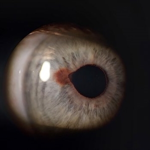
Slit Lamp Photograph of an 88 year old man with an Iris Nevus. Patient presented with DCC 20/60+1. Plan to monitor.
Photographer: Zach Seim
Imaging device: Slit Lamp photography with Samsung Galaxy 7
Condition/keywords: iris, iris nevus, nevus, right eye, slit lamp photo, slit lamp photography
-
 Kissing Serous Choroidal Detachment
Mar 8 2023 by Annaka Gooding
Ultra-widefield fundus photograph of a 73 year old male with a Kissing serous choroidal detachment affecting his right eye. Patient presented at the office following a XEN implant and his vision was sc20/100 PH20/50+1. The physician recommended to start Prednisone treatment.
Photographer: Annaka Gooding
Imaging device: Optos California
Condition/keywords: fundus photography, Kissing Serous Choroidal Detachment, Optos, Right Eye, ultra-wide field imaging
-
 Macula off Retinal Detachment
Jan 23 2024 by Annaka Gooding
Ultra-widefield fundus photograph of an 81-year-old male with a Macula Off Retinal Detachment affecting his right eye. Patient presented at office with complaints of flashes of light for about 2 weeks accompanied by a curtain veil covering inferior visual field. Patient had total vision loss 24 hours prior to visit. His vision was scHM. The physician recommended Retinal Detachment Repair with PPV.
Photographer: Annaka Gooding, CPO
Imaging device: Optos California
Condition/keywords: detachment, fundus photography, macula off retinal detachment, Optos, retinal detachment of the macula, right eye, ultra-wide field imaging
-
 Macula Off Retinal Detachment
Jun 25 2024 by Zach Seim
Optos Fundus photo of a 47 year old female with a Macula Off Retinal Detachment right eye, presenting with loss of nasal visual field. Patient's vision at presentation was DCC 20/100-1. Patient was counseled and decided to proceed with surgery.
Photographer: Zach Seim
Imaging device: OPTOS California
Condition/keywords: macula off retinal detachment, Optos, OPTOS CALIFORNIA, right eye
-
 Macula On Retinal Detachment
Jul 5 2024 by Zach Seim
This is an Optos fundus photo of a 67 year old female with a Macula On Retinal Detachment. Patient presented with VA DCC 20/40-1.
Photographer: Zach Seim
Imaging device: Optos California
Condition/keywords: macula on, Optos, OPTOS CALIFORNIA, right eye
-
 Macular Dystrophy
Oct 25 2023 by Zach Seim
Optos Fundus Autofluorescence of an 84 year old male with Macular Dystrophy. Patient presented with VA of sc CF at 3 feet. Genetic testing was performed to ensure that cause was not genetic.
Photographer: Zach Seim
Imaging device: Optos California
Condition/keywords: Autoflourescence, dystrophy, macular dystrophy, Optos, OPTOS CALIFORNIA, right eye, ultra-wide field imaging
-
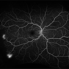
 Neovascularization in Posterior Uveitis
Jul 27 2023 by Zach Seim
An ultra-widefield fluorescein angiogram of a 72 year old male with Posterior Uveitis and Neovascularization affecting the right eye. Patient's vision at the time of the image was Dcc 20/25. Dr. Korot states that the fluorescein angiogram shows patchy leakage throughout both eyes, with peripheral nonperfusion and secondary neovascularization. The patient was asked to get an extensive serological workup in an effort to identify any systemic autoimmune or infectious etiology as the cause for their severe inflammation.
Photographer: Zach Seim
Imaging device: OPTOS California
Condition/keywords: fluorescein angiogram (FA), FLUORESCEIN ANGIOGRAPHY, fluorescein leakage, neovascularization (NV), Optos, OPTOS CALIFORNIA, posterior uveitis, right eye, ultra-wide field imaging, ultra-widefield image
-
 OCT in Subclinical RD in a Case of Retinitis Pigmentosa
May 22 2024 by Tejaswita Verma
OCT of a 6 year old male child showing subretinal fluid with macula off in case of subclinical retinal detachment in retinitis pigmentosa
Photographer: DR. TEJASWITA VERMA
Imaging device: MIRANTE
Condition/keywords: OCT, right eye, subclinical detachment
-
 Proliferative Diabetic Retinopathy with Vitreous Hemorrhage
Sep 28 2022 by Chloe Hanifan
Ultra-widefield pseudo color fundus photograph of a 50-year-old female with Proliferative Diabetic Retinopathy with Vitreous Hemorrhage. The patient's new vitreous hemorrhage symptoms prompted September urgent visit. AntiVEGF was postponed due to chronic health complications with multiple strokes and PRP is recommended in the near future. Her vision was sc20/200-1 PH20/100-2 at the time of her September appointment.
Photographer: Chloe Hanifan
Imaging device: Optos California
Condition/keywords: diabetes, fundus photography, neovascularization, Optos, proliferative diabetic retinopathy (PDR), pseudocolor, right eye, ULTRA WIDE FIELD, vitreous hemorrhage
-
 Proliferative Sickle Cell Retinopathy
Feb 1 2023 by Olivia Rainey
Ultra-widefield fluorescein angiography of a 25-year old male with Proliferative Sickle Cell Retinopathy affecting his right eye. Patient stated that he was born with Sickle disease (SC), and has yearly eye exams. He noted no vision concerns over the last year but has typically experienced sickle attacks about 1-2 per year. The physician noted that the fluorescein obtained showed peripheral nonperfusion affecting the patient's nasal and temporal retina as well as neovascularization affecting his left eye more than his right. He recommended pan retinal photocoagulation in his left eye for his temporal and nasal retina, as as well as his right eye following.
Photographer: Olivia Rainey, OCT-C, COA
Imaging device: Optos California
Condition/keywords: early phase, fluorescein angiogram (FA), fluorescein leakage, neovascularization (NV), non-perfusion, proliferative retinopathy, right eye, sickle cell retinopathy, ultra-wide field imaging, ultra-widefield image
-
 Prominent Long Ciliary Nerve
Jan 25 2022 by Kachelle Brown
Ultra-wide field photograph of a 48-year-old female with a prominent long ciliary nerve. Patient presented asymptomatic, and was referred for a macula on retinal detachment. Patient was diagnosed with high myopia and a posterior vitreous detachment, and the physician discussed increased risk of floaters, myopic degeneration and retinal detachment associated with high myopia. -24.00 prior to cataract surgery OU per patient.
Photographer: Kachelle Brown
Imaging device: Optos California
Condition/keywords: fundus photograph, high myopia, long ciliary nerve, optos, right eye, ultra-widefield image
-
 Scalloped Choroidal Atrophy
Jan 8 2024 by Zach Seim
An ultra-widefield fluorescein angiogram of a 90 year old female with Scalloped Choroidal Atrophy affecting both eyes. Patient's vision at the time of the image was Dcc 20/40 OD. Genetic test pending.
Photographer: Zach Seim
Imaging device: OPTOS California
Condition/keywords: atrophy, choroidal atrophy, fluorescein angiogram (FA), Fluorescein angiography, optic nerve, OPTOS CALIFORNIA, retina, right eye, ultra-wide field imaging
-
 Sturge-Weber Syndrome
Nov 17 2023 by Zach Seim
Topcon photo of a 37 year old female with Sturge-Weber syndrome affecting OU. Patient presents with prominent episcleral vasculature and DCC 20/20 VA OU. Plan to monitor.
Photographer: Zach Seim
Imaging device: Topcon 50DX
Condition/keywords: bilateral, external, external photography, left eye, right eye, Sturge-Weber syndrome, Topcon
-
 Vitreomacular Traction
Jun 15 2022 by Zach Seim
Optical Coherence Tomography (OCT) of a 69 year old male with Vitreomacular Traction affecting his right eye. Patient was referred to this office for Choroidal Melanoma in his right eye in May 2021. The patient was treated with Brachytherapy in July 2021 and this OCT was taken at a follow-up appointment in May 2022. Patient's vision was 20/30-2 at the time this OCT was taken. Patient states that his vision was better since his last visit, and that he sees floaters occasionally.
Photographer: Zach Seim
Imaging device: Heidelberg Spectralis
Condition/keywords: heidelberg spectralis, OD, optical coherence tomography (OCT), right eye, subretinal fluid, vitreomacular adhesion, vitreomacular interface disorders, vitreomacular traction (VMT)
-
 A Large Break at the Posterior Pole With RD With PVR (S/p Old Blunt Trauma)
Jan 16 2025 by Anand Temkar
Right eye central fundus color photo of a 10 year old kid who noticed diminution of vision in right eye since a month. We can see the large break at the posterior pole with rolled up margins associated with retinal detachment and PVR changes.
Photographer: Dr.Anand Temkar- Retina Foundation, Ahmedabad
Imaging device: Mirante
Condition/keywords: Posterior pole break, proliferative vitreoretinopathy (PVR), Retinal Detachment
-
 A Large Break at the Posterior Pole With RD With PVR (S/p Old Blunt Trauma)
Jan 16 2025 by Anand Temkar
Right eye widefield fundus color photo of a 10 year old kid who noticed diminution of vision in right eye since a month. We can see the large break at the posterior pole with rolled up margins associated with retinal detachment and PVR changes.
Photographer: Dr.Anand Temkar- Retina Foundation, Ahmedabad
Imaging device: Mirante
Condition/keywords: posterior pole break, proliferative vitreoretinopathy (PVR), Retinal Detachment
-
---thumb.JPG/image-square;max$300,300.ImageHandler) Acute myeloid leukemia
Acute myeloid leukemia
Dec 9 2012 by Mallika Goyal, MD
Right eye of a 21-year-old gentleman with acute myeloid leukemia who is undergoing chemotherapy and has low platelet counts (17,000) shows multiple pre-retinal haemorrhages. Other eye has similar picture. There is no vascular occlusion or inflammation. Visual prognosis remains good with spontaneous resolution expected over few weeks.
Photographer: Mallika Goyal, MD, Apollo Health City, Hyderabad, India
Condition/keywords: preretinal hemorrhage
 Loading…