Search results (26 results)
-
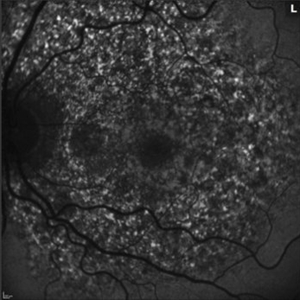
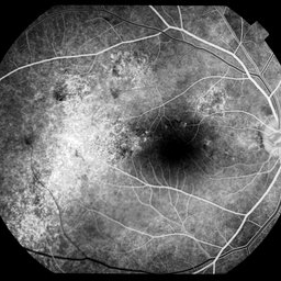 RPE Mottling
Jul 7 2025 by Moazzam Parvez
Fundus fluorescein image of a 62 year old gentleman in the early phase showing diffuse RPE mottling in the temporal aspect of the arcade.
Photographer: Moazzam Parvez
Imaging device: Heidelberg Spectralis
Condition/keywords: FA early phase, FFA, RPE mottling
-
---thumb.jpg/image-square;max$300,300.ImageHandler) Pattern Dystrophy
Pattern Dystrophy
Aug 9 2013 by From the Collections of Thomas M. Aaberg, MD and Thomas M. Aaberg Jr., MD
RPE mottling, right eye.
Condition/keywords: pattern macular dystrophy, retinal pigment epithelium
-
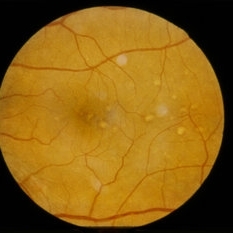
 Congenital Nyctalopia
Feb 20 2013 by From the Collections of Thomas M. Aaberg, MD and Thomas M. Aaberg Jr., MD
Color photo of the periphery showing RPE mottling, clumping, and small multiple yellow deposits.
Condition/keywords: color photo, congenital nyctalopia, retinal pigment epithelium
-
---thumb.jpg/image-square;max$300,300.ImageHandler) Congenital Nyctalopia
Congenital Nyctalopia
Feb 20 2013 by From the Collections of Thomas M. Aaberg, MD and Thomas M. Aaberg Jr., MD
Color photo of the optic nerve showing mild peripaillary RPE mottling.
Condition/keywords: color photo, congenital nyctalopia, retinal pigment epithelium
-
---thumb.jpg/image-square;max$300,300.ImageHandler) Congenital Nyctalopia
Congenital Nyctalopia
Feb 20 2013 by From the Collections of Thomas M. Aaberg, MD and Thomas M. Aaberg Jr., MD
Color photo of the periphery showing RPE mottling, clumping, and small multiple yellow deposits.
Condition/keywords: color photo, congenital nyctalopia, retinal pigment epithelium
-
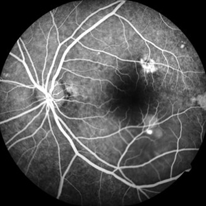 CSR FA
May 15 2021 by Deepak Bhojwani, MS
Fundus fluoroscein angiography image of a 38-year-old gentlemen with features of chronic CSR with multiple old areas of RPE mottling and staining. Also note there is a single active leakage site along inferotemporal arcade.
Photographer: Deepak Bhojwani
Condition/keywords: central serous retinopathy (CSR)
-
 LCA Type 2
Apr 10 2025 by Joshua Friedman
LCA Type 2 (RPE65) showing characteristic hypoautofluorescence and retinal thinning. 8F with best corrected visual acuity of 20/400 (OD) and 20/150 (OS). Small white intraretinal spots and RPE mottling are visible on color fundus photography. Blue light autofluorescence reveals near-complete loss of signal, while OCT demonstrates widespread outer retinal thinning.
Photographer: Stephen Tsang, MD, PhD
Condition/keywords: Leber Congenital Amaurosis
-
 Myopic Maculoscheisis
Sep 11 2013 by Jason S. Calhoun
Fluorescence angiography and fundus photography shows RPE mottling and staphyloma in the right eye. VA is 20/50, right eye. No subretinal fluid found.
Photographer: Jason S. Calhoun, Department of Ophthalmology, Mayo Clinic Jacksonville, Florida
Imaging device: TOPCON TRC 50-EX
Condition/keywords: myopia, tilted disc
-
---thumb.jpg/image-square;max$300,300.ImageHandler) Non-Staining CME
Non-Staining CME
Feb 20 2013 by From the Collections of Thomas M. Aaberg, MD and Thomas M. Aaberg Jr., MD
FA of OD with RPE mottling at macula with no late leakage (possibly an eye with RP).
Condition/keywords: non-staining cystoid macular edema (CME)
-
 Optic Disc Pit
Dec 11 2014 by H. Michael Lambert, MD
Fluorescein angiogram of RE with ON pit and macular detachment with RPE mottling.
Condition/keywords: optic disc pit
-
---thumb.jpg/image-square;max$300,300.ImageHandler) Pattern Dystrophy
Pattern Dystrophy
Aug 12 2013 by From the Collections of Thomas M. Aaberg, MD and Thomas M. Aaberg Jr., MD
FA with RPE mottling.
Condition/keywords: pattern macular dystrophy, retinal pigment epithelium
-
---thumb.jpg/image-square;max$300,300.ImageHandler) Pattern Dystrophy
Pattern Dystrophy
Aug 12 2013 by From the Collections of Thomas M. Aaberg, MD and Thomas M. Aaberg Jr., MD
FA with RPE mottling.
Condition/keywords: pattern macular dystrophy, retinal pigment epithelium
-
 Pentosan Maculopathy Autofluorescence Left
Oct 25 2020 by Thomas A. Ciulla, MD, MBA, FASRS
56-year-old woman with prior long term use of pentosan for cystitis. She self discontinued the pentosan 1 year ago. Imaging shows patch RPE atrophy OU with a ¾ DA patch of geo atrophy OS. Autofluorescence shows punctate hyperautoflouresence and FA shows staining of RPE mottling with reticular features.
Condition/keywords: pentosan sulfate
-
 Pentosan Maculopathy Autofluorescence Right
Oct 25 2020 by Thomas A. Ciulla, MD, MBA, FASRS
56-year-old woman with prior long term use of pentosan for cystitis. She self discontinued the pentosan 1 year ago. Imaging shows patch RPE atrophy OU with a ¾ DA patch of geo atrophy OS. Autofluorescence shows punctate hyperautoflouresence and FA shows staining of RPE mottling with reticular features.
Condition/keywords: pentosan sulfate
-
 Pentosan Maculopathy Color Fundus Photo Left
Oct 25 2020 by Thomas A. Ciulla, MD, MBA, FASRS
56-year-old woman with prior long term use of pentosan for cystitis. She self discontinued the pentosan 1 year ago. Imaging shows patch RPE atrophy OU with a ¾ DA patch of geo atrophy OS. Autofluorescence shows punctate hyperautoflouresence and FA shows staining of RPE mottling with reticular features.
Condition/keywords: pentosan sulfate
-
 Pentosan Maculopathy Color Fundus Photo Right
Oct 25 2020 by Thomas A. Ciulla, MD, MBA, FASRS
56-year-old woman with prior long term use of pentosan for cystitis. She self discontinued the pentosan 1 year ago. Imaging shows patch RPE atrophy OU with a ¾ DA patch of geo atrophy OS. Autofluorescence shows punctate hyperautoflouresence and FA shows staining of RPE mottling with reticular features.
Condition/keywords: pentosan sulfate
-
 Pentosan Maculopathy Fluorescein Angiogram Left
Oct 25 2020 by Thomas A. Ciulla, MD, MBA, FASRS
56-year-old woman with prior long term use of pentosan for cystitis. She self discontinued the pentosan 1 year ago. Imaging shows patch RPE atrophy OU with a ¾ DA patch of geo atrophy OS. Autofluorescence shows punctate hyperautoflouresence and FA shows staining of RPE mottling with reticular features.
Condition/keywords: pentosan sulfate
-
 Pentosan Maculopathy Fluorescein Angiogram Right
Oct 25 2020 by Thomas A. Ciulla, MD, MBA, FASRS
56-year-old woman with prior long term use of pentosan for cystitis. She self discontinued the pentosan 1 year ago. Imaging shows patch RPE atrophy OU with a ¾ DA patch of geo atrophy OS. Autofluorescence shows punctate hyperautoflouresence and FA shows staining of RPE mottling with reticular features.
Condition/keywords: pentosan sulfate
-
 Pentosan Maculopathy OCT Left
Oct 25 2020 by Thomas A. Ciulla, MD, MBA, FASRS
56-year-old woman with prior long term use of pentosan for cystitis. She self discontinued the pentosan 1 year ago. Imaging shows patch RPE atrophy OU with a ¾ DA patch of geo atrophy OS. Autofluorescence shows punctate hyperautoflouresence and FA shows staining of RPE mottling with reticular features.
Condition/keywords: pentosan sulfate
-
 Pentosan Maculopathy OCT Right
Oct 25 2020 by Thomas A. Ciulla, MD, MBA, FASRS
56-year-old woman with prior long term use of pentosan for cystitis. She self discontinued the pentosan 1 year ago. Imaging shows patch RPE atrophy OU with a ¾ DA patch of geo atrophy OS. Autofluorescence shows punctate hyperautoflouresence and FA shows staining of RPE mottling with reticular features.
Condition/keywords: pentosan sulfate
-
 Pentosan Maculopathy Red Free Photo Left
Oct 25 2020 by Thomas A. Ciulla, MD, MBA, FASRS
56-year-old woman with prior long term use of pentosan for cystitis. She self discontinued the pentosan 1 year ago. Imaging shows patch RPE atrophy OU with a ¾ DA patch of geo atrophy OS. Autofluorescence shows punctate hyperautoflouresence and FA shows staining of RPE mottling with reticular features.
Condition/keywords: pentosan sulfate
-
 Pentosan Maculopathy Red Free Photo Right
Oct 25 2020 by Thomas A. Ciulla, MD, MBA, FASRS
56-year-old woman with prior long term use of pentosan for cystitis. She self discontinued the pentosan 1 year ago. Imaging shows patch RPE atrophy OU with a ¾ DA patch of geo atrophy OS. Autofluorescence shows punctate hyperautoflouresence and FA shows staining of RPE mottling with reticular features.
Condition/keywords: pentosan sulfate
-
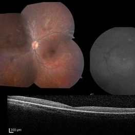
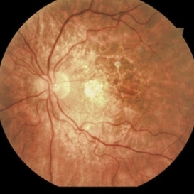
 Stargardt's Disease
Feb 12 2015 by H. Michael Lambert, MD
Right eye, RPE mottling at macular with central and paracentral flecks.
Condition/keywords: macular degeneration, Stargardt disease
 Loading…