Search results (9 results)
-
 Imaging Feeder Vessels on OCT-A in a case of Retinal Angiomatous Proliferation
Feb 24 2023 by Dhaivat Shah
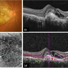
A 58-year-old male patient, a chronic smoker, came to our OPD with complaints of a diminution of vision in the right eye (BCVA: 2/60). On examination, the following findings were observed in the patient. On fundus examination (Image 1a) - Large areas of exudation with multiple superficial and deep hemorrhages at the macula were noted. On SD-OCT imaging (Image 1b) - Multiple intraretinal spaces were seen along with shallow subretinal fluid and hyperreflective dots (indicative of phagocytosed photoreceptors). On the foveal area- a hyperreflective membrane was noted which seemed to dip down and establish a retino-choroidal anastomosis. On OCT-A imaging (Image 1c) - In the ORCC complex, the neovascularization frown, correlating with the membrane complex on the fundus and structural OCT, was visible which was noted to be supplied by small feeder vessels coming from the superior aspect of the fovea. On OCT-A blood flow analysis imaging (Image 1d) - The blood flow analysis showed increased blood flow signals at the level of the membrane (indicative of increased color signal). Based on the findings of the above investigations and clinical examination, the patient was diagnosed with a Case of CNVM Type 3, also described as RAP, and was managed by Anti VEGF injections. This condition usually requires more injections as compared to Type 1 and 2 CNVMs, and the visual prognosis is guarded. Hence, it's very important to counsel the patient before initiating the treatment that the treatment would be long-term and the aim would be preservation of existing vision.
Photographer: Choithram Netralaya, Indore
Condition/keywords: feeder vessel, OCTA, RAP lesion
-
 Retinal Angiomatos Proliferans
Jun 10 2020 by Manish Nagpal, MD, FRCS (UK), FASRS
RAP lesion
Photographer: Gayathri Mohan, Retina Foundation
Imaging device: NIDEK SLO MIRANTE
Condition/keywords: RAP lesion
-
 Retinal Angiomatous Proliferation
Sep 10 2018 by Gabriela Lopezcarasa Hernandez, MD
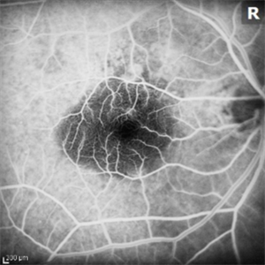
75-year-old patient with decrease in visual acuity right eye with metamorphopsia, in the FA and ICG we can see a RAP lesion.
Photographer: Azucena Rios
Imaging device: Heidelberg Spectralis
Condition/keywords: FA mid phase, indocyanine green (ICG) angiography, RAP lesion, retinal angiomatous proliferation (RAP)
-
 RAP lesions
Sep 29 2014 by Thomas A. Ciulla, MD, MBA, FASRS
OCT of an 81-year-old man revealing several RAP lesions superior to fovea.
Photographer: Stuart Alfred CRA
Condition/keywords: choroidal neovascular membrane (CNVM), neovascular age-related macular degeneration (AMD), retinal angiomatous proliferation (RAP), wet age-related macular degeneration (wet AMD)
-
 RAP lesions
Sep 29 2014 by Thomas A. Ciulla, MD, MBA, FASRS
Late frame fluorescein angiogram of an 81-year-old man revealing several RAP lesions superior to fovea.
Photographer: Stuart Alfred CRA
Condition/keywords: choroidal neovascular membrane (CNVM), neovascular age-related macular degeneration (AMD), retinal angiomatous proliferation (RAP), wet age-related macular degeneration (wet AMD)
-
 RAP lesions
Sep 29 2014 by Thomas A. Ciulla, MD, MBA, FASRS
Fluorescein angiogram of an 81-year-old man revealing several RAP lesions superior to fovea.
Photographer: Stuart Alfred CRA
Condition/keywords: choroidal neovascular membrane (CNVM), neovascular age-related macular degeneration (AMD), retinal angiomatous proliferation (RAP), wet age-related macular degeneration (wet AMD)
-
 RAP Lesions
Sep 29 2014 by Thomas A. Ciulla, MD, MBA, FASRS
Fluorescein angiogram of an 81-year-old man revealing several RAP lesions superior to fovea.
Photographer: Stuart Alfred CRA
Condition/keywords: choroidal neovascular membrane (CNVM), neovascular age-related macular degeneration (AMD), retinal angiomatous proliferation (RAP), wet age-related macular degeneration (wet AMD)
-
 RAP lesions
Sep 29 2014 by Thomas A. Ciulla, MD, MBA, FASRS
Fluorescein angiogram of an 81-year-old man revealing several RAP lesions superior to fovea.
Photographer: Stuart Alfred CRA
Condition/keywords: choroidal neovascular membrane (CNVM), neovascular age-related macular degeneration (AMD), retinal angiomatous proliferation (RAP), wet age-related macular degeneration (wet AMD)
-
 Stereo View of Retinal Angiomatous Proliferation in Age-Related Macular Degeneration
Jan 21 2016 by James B. Soque, CRA, OCT-C, COA, FOPS
Stereo pair of 75-year-old white male with classic SRN with RAP lesion of right eye, actively receiving anti-VEGF treatment. 50 Degrees, no mag, L and R stereo pair. Single View of OD also visible in this case.
Condition/keywords: age-related macular degeneration (AMD), anti-VEGF, retinal angiomatous proliferation (RAP), stereo pair, subretinal neovascularization (SRNV)
 Loading…