Search results (37 results)
-
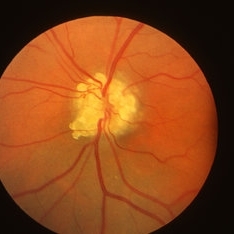
 Optic Nerve Head Drusen
Dec 10 2012 by Yale L. Fisher, MD
Another movie from Dr. Yale Fisher and the OphthalmicEdge.org Contact B-scan ultrasonography can be of great help in diagnosing buried drusen of the optic nerve head. When drusen are buried deep in the nerve and invisible to white light, ultrasound has proved to be diagnostically helpful. Drusen of the nerve head are excellent reflectors of sound (probably due to the presence of calcification). Drusen themselves have an extensive shadowing effect, but since we are already shooting through the optic nerve shadow, much of this effect is not evident. They stand out like small pebbles on the head of the optic nerve and their characteristic hyper-reflective signal persist at the lowest decibel levels of gain. Also in this eye, scattered vitreous opacities of very low reflectivity are visible, which may be related to advancing age. In younger eyes, the clear vitreous body generally produces no echoes.
Condition/keywords: video
-
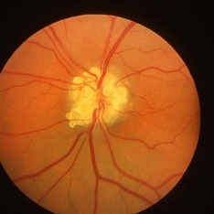
 Optic Nerve Head Drusen
Oct 2 2013 by Jerald A. Bovino, MD
There are optic nerve head drusen. These are also visualized on fundus autofluorescence and B-scan ultrasonography.
Condition/keywords: optic nerve drusen
-
 Optic Nerve Head Drusen
Oct 2 2013 by Jerald A. Bovino, MD
There are optic nerve head drusen. These are also visualized on fundus autofluorescence and B-scan ultrasonography.
Condition/keywords: optic nerve drusen
-
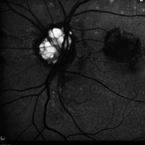
 Optic Nerve Head Drusen
Sep 10 2015 by Mariam A Al-Feky, MD
Right eye, 30-year-old obese female patient with BCVA 0.8 OU presenting with severe headache of 1 month duration. Ant seg: NAD OU, post. segment: disc edema OU, MRI brain is normal. ONH drusen is the diagnosis actually, with late staining in FFA (without early hyperfluorescent telangectatic disc capillaries as in caes of papillaedema), well delineated in the ONH map on the Heidelberg machine, and could be detected as a hypereflective material below the nerve fiber layer on the line scan. N.B. Burried ONH drusen don't autofluoresce.
Imaging device: Fundus camera
Condition/keywords: optic nerve head
-
 Optic Nerve Head Drusen
Sep 10 2015 by Mariam A Al-Feky, MD
Left eye 30-year-old obese female patient with BCVA 0.8 OU presenting with severe headache of 1 month duration Ant seg: NAD OU, post. segment: disc edema OU MRI brain is normal ONH drusen is the diagnosis actually, with late staining in FFA (without early hyperfluorescent telangectatic disc capillaries as in cases of papillaedema), well delineated in the ONH map on the Heidelberg machine, and could be detected as a hypereflective material below the nerve fiber layer on the line scan. N.B. Burried ONH drusen don't autofluoresce.
Imaging device: Fundus camera
Condition/keywords: drusen, optic nerve head
-
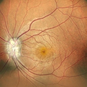
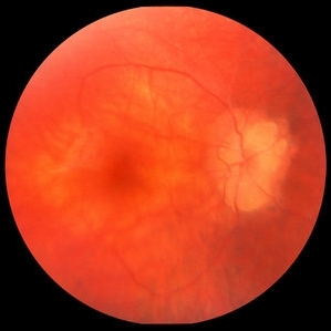
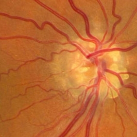 Optic Nerve Head Drusen
Feb 12 2015 by Timothy S Fuller, MD
Fundus photograph of a 34-year-old woman with striking, asymptomatic nerve head drusen.
Photographer: Nick Hesse, Texas Retina Associates
Condition/keywords: drusen of optic disc
-
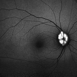
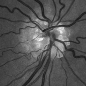 Optic Nerve Head Drusen
Feb 12 2015 by Timothy S Fuller, MD
Fundus autofluorescence image of a 34-year-old woman with striking, asymptomatic nerve head drusen.
Photographer: Nick Hesse, Texas Retina Associates
Imaging device: Heidelberg Spectralis
Condition/keywords: drusen of optic disc
-
 Optic Nerve Head Drusen
Feb 12 2015 by Timothy S Fuller, MD
Fundus photograph of a 34-year-old woman with striking, asymptomatic optic nerve head drusen.
Photographer: Nice Hesse, Texas Retina Associates
Condition/keywords: drusen of optic disc
-
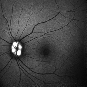
 Optic Nerve Head Drusen
Feb 12 2015 by Timothy S Fuller, MD
Fundus autofluorescence image of a 34-year-old woman with striking, asymptomatic optic nerve head drusen.
Photographer: Nice Hesse, Texas Retina Associates
Imaging device: Heidelberg Spectralis
Condition/keywords: drusen of optic disc
-
 Optic Nerve Head Drusen
Feb 9 2018 by Olivia Rainey
Fundus autofluorescence of a 49-year-old female with optic nerve head drusen affecting her left eye. The patient has pseudoxanthoma elasticum with choroidal neovascularization and has been receiving anti-VEGF treatment for many years.
Photographer: Olivia Rainey
Imaging device: Heidelberg Spectralis
Condition/keywords: 30 degrees, anti-VEGF, choroidal neovascularization (CNV), fundus autofluorescence (FAF), Heidelburg Spectralis, left eye, optic disc, optic nerve drusen, pseudoxanthoma elasticum (PXE)
-
 Optic Nerve Head Drusen
Feb 20 2013 by From the Collections of Thomas M. Aaberg, MD and Thomas M. Aaberg Jr., MD
No history; Color and FA; probable infero-temporal visual field defect.
Condition/keywords: optic nerve drusen
-
 Optic Nerve Head Drusen
Feb 20 2013 by From the Collections of Thomas M. Aaberg, MD and Thomas M. Aaberg Jr., MD
No history; Color and FA; probable superior altitudinal visual field defect
Condition/keywords: optic nerve drusen
-
 Optic Nerve Head Drusen
Feb 20 2013 by From the Collections of Thomas M. Aaberg, MD and Thomas M. Aaberg Jr., MD
No history; Color and FA.
Condition/keywords: optic nerve drusen
-
 Optic Nerve Head Drusen
Feb 20 2013 by From the Collections of Thomas M. Aaberg, MD and Thomas M. Aaberg Jr., MD
No history, FA, probable superior altitudinal visual field defect.
Condition/keywords: optic nerve drusen
-
---thumb.jpg/image-square;max$300,300.ImageHandler) Optic Nerve Head Drusen
Optic Nerve Head Drusen
Feb 20 2013 by From the Collections of Thomas M. Aaberg, MD and Thomas M. Aaberg Jr., MD
drusen
Condition/keywords: optic nerve drusen
-
---thumb.jpg/image-square;max$300,300.ImageHandler) Optic Nerve Head Drusen
Optic Nerve Head Drusen
Feb 20 2013 by From the Collections of Thomas M. Aaberg, MD and Thomas M. Aaberg Jr., MD
FA of the right optic nerve demonstrating hyperfluorescence
-
 Optic nerve head drusen
Dec 26 2022 by Vaidehi Sathaye
FAF photograph of RE of a 32 year old female with Optic nerve head drusen
Photographer: Dr. Vaidehi Sathaye
Imaging device: Mirante
Condition/keywords: drusen of optic disc
-
 Optic nerve head drusen
Dec 26 2022 by Vaidehi Sathaye
FAF photograph of LE of a 32 year old female with Optic nerve head drusen
Photographer: Dr. Vaidehi Sathaye
Imaging device: Mirante
Condition/keywords: drusen of optic disc
-
 Optic nerve head drusen
Dec 26 2022 by Vaidehi Sathaye
Fundus Photograph of LE of a 32 year old female with Optic nerve head drusen
Photographer: Dr. Vaidehi Sathaye
Imaging device: Mirante
Condition/keywords: drusen of optic disc
-
 Optic nerve head drusen
Dec 26 2022 by Vaidehi Sathaye
Fundus Photograph of RE of a 32 year old female with Optic nerve head drusen
Photographer: Dr. Vaidehi Sathaye
Imaging device: Mirante
Condition/keywords: drusen of optic disc
-
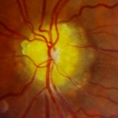
 Optic Nerve Head Drusen
Oct 16 2012 by Jeffrey G. Gross, MD, FASRS
Optic nerve head drusen.
Condition/keywords: optic nerve drusen
-
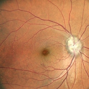
 Optic nerve head drusen
Sep 14 2023 by Ben Serar
Fundus photograph showing yellowish-white nodules at the optic disc causing blurring of the disc margins in a case of Optic nerve head drusen.
Condition/keywords: Optic nerve head drusen
-
 Optic nerve head drusen
Sep 14 2023 by Ben Serar
Fundus photograph showing a yellowish-white nodule at the superior margin of the optic disc in a case of Optic nerve head drusen.
Condition/keywords: Optic nerve head drusen
-
---thumb.jpg/image-square;max$300,300.ImageHandler) Drusen
Drusen
-
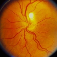
 Optic Nerve Head Drusen - Fundus Image
Oct 5 2013 by Roy Schwartz, MD
Optice nerve head drusen in a right eye, fundus image.
Photographer: Galit Yair-Pur
Condition/keywords: disc drusen, drusen of optic disc, optic nerve drusen, red-free
 Loading…