Search results (7 results)
-
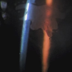 Ink Blot Epithelial Ingrowth Post-LASIK Refractive Surgery
Jun 29 2024 by Luai Abu-Ismail, MD
Anterior segment photo of a 45-year-old female patient presented 12-year post-LASIK surgery.
Photographer: Dr. Luai Abu-Ismail, Ophthalmology Department, Islamic Hospital.
Imaging device: Slit lamp biomicroscope photo taken by Smart phone camera.
Condition/keywords: complication, cornea, corneal scars and opacities, epithelial ingrowth, LASIK, LASIK FLAP, refractive surgery
-
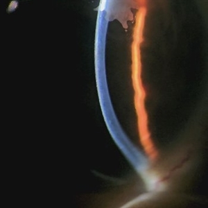 Ink Blot Epithelial Ingrowth Post-LASIK Refractive Surgery
Jun 29 2024 by Luai Abu-Ismail, MD
Anterior segment photo of a 45-year-old female patient presented 12-year post-LASIK surgery.
Photographer: Dr. Luai Abu-Ismail, Ophthalmology Department, Islamic Hospital.
Imaging device: Slit lamp biomicroscope photo taken by Smart phone camera.
Condition/keywords: cornea, corneal scars and opacities, flap, LASIK
-
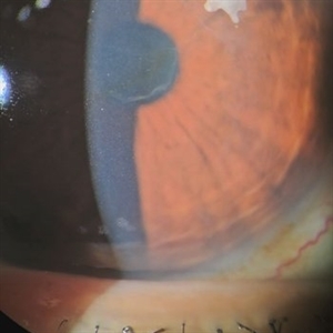 Ink Blot Epithelial Ingrowth Post-LASIK Refractive Surgery
Jun 29 2024 by Luai Abu-Ismail, MD
Anterior segment photo of a 45-year-old female patient presented 12-year post-LASIK surgery.
Photographer: Dr. Luai Abu-Ismail, Ophthalmology Department, Islamic Hospital.
Imaging device: Slit lamp biomicroscope photo taken by Smart phone camera.
Condition/keywords: cornea, corneal scars and opacities, flap, LASIK
-
 Radial Keratotomy (RK)
Jul 13 2013 by Jason S. Calhoun
Normal slit lamp photo that shows s/p RK scars.
Photographer: Jason S. Calhoun, Department of Ophthalmology, Mayo Clinic Jacksonville, Florida
Imaging device: TOPCON D-90 SL NIKON CAMERA
Condition/keywords: LASIK
-
---thumb.JPG/image-square;max$300,300.ImageHandler) Radial Keratotomy (RK) Scars
Radial Keratotomy (RK) Scars
Jul 13 2013 by Jason S. Calhoun
Normal slit lamp photo that shows s/p RK scars.
Photographer: Jason S. Calhoun, Department of Ophthalmology, Mayo Clinic Jacksonville, Florida
Imaging device: TOPCON D-90 SL NIKON CAMERA
Condition/keywords: LASIK
-
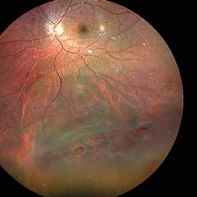 Inferior retinal detachment with lattice and holes
May 31 2023 by Aditya S Kelkar, MS, FRCS, FASRS,FRCOphth
Importance of dilated retina check up before Lasik surgery can't be better demonstrated...patient totally asymptomatic came for Lasik opinion and has inferior retinal detachment with lattice and holes, sparing the macula
Photographer: Dr. Sahil Wagh , National Institute of Opthalmology, Pune , India
Imaging device: Zeiss Clarus 500
Condition/keywords: inferior retinal detachment
-
 Retinal Detachment with Proliferative Vitreoretinopathy
Mar 20 2014 by Min Kim, MD, PhD, MBA, FASRS
Wide field fundus photograph of a 59-year-old male with chronic total RD and PVR, with multiple retinal breaks that developed a few months after LASIK surgery.
Photographer: Young Duk Bae, Yonsei University, Gangnam Severance Hospital
Imaging device: Wide field fundus photography, Optomap
Condition/keywords: proliferative vitreoretinopathy (PVR), retinal detachment with retinal defect
 Loading…