Search results (26 results)
-
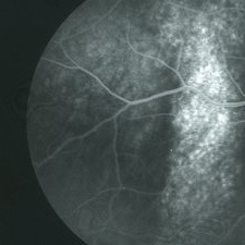
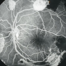
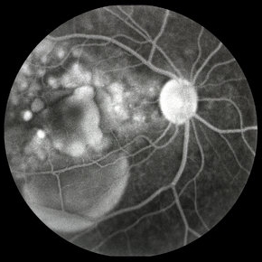
 Bilateral Central Serous Retinopathy
Mar 26 2019 by Gary R. Cook, MD, FACS
Mid-phase fluorescein angiogram frame of a pinpoint leak in the temporal macula OS of a 37-year-old white male with bilateral central serous retinopathy; VA = 20/15+3.
Imaging device: Topcon VT-50
Condition/keywords: central serous retinopathy (CSR), FA mid phase, fluorescein angiogram (FA)
-
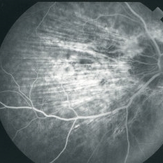 C-R Folds
Mar 26 2019 by Gary R. Cook, MD, FACS
Mid-phase FA image of the right eye of a white male with bilateral C-R folds showing alternating hyper- and hypofluorescent bands.
Imaging device: Topcon VT-50
Condition/keywords: bilateral chorioretinal folds, chorioretinal fold, FA mid phase, fluorescein angiogram (FA)
-
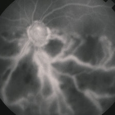
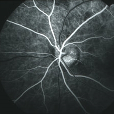 Capillary Hemangioma
Apr 1 2019 by Gary R. Cook, MD, FACS
Mid-phase (laminar venous phase) fluorescein angiogram image of a capillary hemangioma of the optic disc OS showing delayed filling and relative hypofluorescence in the area of the hemangioma on the superior aspect in a 28-year-old white female
Imaging device: Topcon VT-50
Condition/keywords: FA mid phase, fluorescein angiogram (FA), hemangioma
-
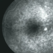
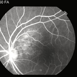
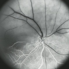
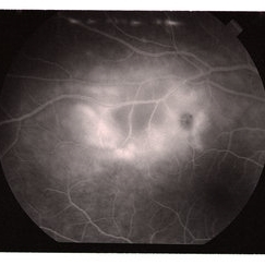
 Central Serous Retinopathy
Mar 26 2019 by Gary R. Cook, MD, FACS
Venous phase of FA of left eye of 45-year-old white male with CSR showing multiple RPE abnormalities and a depigmented RPE track in temporal macula going inferiorly; VA= 20/30.
Imaging device: Topcon VT-50
Condition/keywords: central serous retinopathy (CSR), FA mid phase, fluorescein angiogram (FA)
-
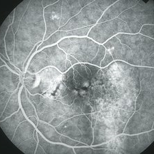
 Central Serous Retinopathy
Mar 26 2019 by Gary R. Cook, MD, FACS
Mid-phase FA frame of right eye of a 45-year-old white male with a history of bilateral CSR showing stippled hyperfluorescence in an area of an RPE track beneath the inferotemporal arcade OD; no active dye leakage is present; VA = 20/200.
Imaging device: Topcon VT-50
Condition/keywords: central serous retinopathy (CSR), FA mid phase, fluorescein angiogram (FA)
-
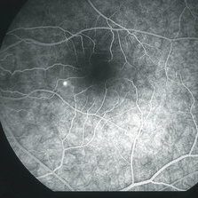 Central Serous Retinopathy
Mar 26 2019 by Gary R. Cook, MD, FACS
32 year old white female with acute CSR OS; early venous filling phase of FA showing a pinpoint leak in inferonasal macula; VA = 20/30-2.
Imaging device: Topcon VT-50
Condition/keywords: central serous retinopathy (CSR), FA mid phase, fluorescein angiogram (FA), fluorescein leakage
-
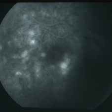
 Choroidal Hemangioma
Sep 15 2014 by Thomas A. Ciulla, MD, MBA, FASRS
Mid-phase fluorescein angiography reveals a well-circumscribed coarse hyperfluorescent vascular pattern within the lesion.
Photographer: Charlotte Harris
Condition/keywords: choroidal hemangioma, choroidal tumor, FA mid phase
-
 Choroidal Hemangioma
Sep 15 2014 by Thomas A. Ciulla, MD, MBA, FASRS
Mid-phase fluorescein angiography reveals a well-circumscribed coarse hyperfluorescent vascular pattern within the lesion.
Photographer: Charlotte Harris
Condition/keywords: choroidal hemangioma, choroidal tumor, FA mid phase
-
 Choroidal Hemangioma
Sep 15 2014 by Thomas A. Ciulla, MD, MBA, FASRS
Mid-phase fluorescein angiography reveals a well-circumscribed coarse hyperfluorescent vascular pattern within the lesion.
Photographer: Charlotte Harris
Condition/keywords: choroidal hemangioma, choroidal tumor, FA mid phase
-
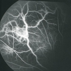 Eales Disease
Apr 1 2019 by Gary R. Cook, MD, FACS
Mid-phase fluorescein angiogram image of the left eye of a 23-year-old Vietnamese female with Eales Disease showing the retinal vascular abnormalities, capillary loss, and a focus of NVE; V.A.= 20/25-2.
Imaging device: Topcon VT-50
Condition/keywords: Eales disease, FA mid phase, fluorescein angiogram (FA), neovascularization elsewhere (NVE)
-
 Eales Disease
Apr 1 2019 by Gary R. Cook, MD, FACS
Mid-phase fluorescein angiogram frame of the left eye of a 23-year-old Vietnamese female with Eales Disease showing multiple areas of NVE and areas of capillary loss and nonperfusion OS.
Imaging device: Topcon VT-50
Condition/keywords: Eales disease, FA mid phase, fluorescein angiogram (FA), neovascularization elsewhere (NVE)
-
 Fundus Fluorescein Angiography of Choroidal Metastases
Jan 18 2020 by Vishal Agrawal, MD, FRCS,FACS,FASRS
Left eye FFA montage of a 55-year-old female with choroidal metastases with the primary being breast carcinoma. The right eye had exudative retinal detachment . Note the pin point leaks at the border of the 2 lesions.
Photographer: Dr Vishal Agrawal MD,FRCS
Imaging device: Zeiss
Condition/keywords: breast cancer, FA mid phase, metastatic lesion
-
 Hemi-CRAO
Mar 26 2019 by Gary R. Cook, MD, FACS
Mid-phase (laminar venous return) fluorescein angiogram image of an embolic superior hemi-CRAO showing marked delay in filling of the superior retinal arteriolar and venous vasculature and total loss of the retinal capillary bed in the superior hemisphere OD.
Condition/keywords: capillary closure, capillary nonperfusion, central retinal artery occlusion (CRAO), FA mid phase, fluorescein angiogram (FA)
-
 Hemi-CRVO
Jun 4 2019 by Gary R. Cook, MD, FACS
Mid-phase FA image of 78-year-old African American female patient with COAG and a very ischemic inferior hemi-CRVO showing loss of almost all of the capillary bed and staining of the veins in the lower hemisphere of the left eye; V.A. = HM 1 ft.
Imaging device: Topcon VT-50
Condition/keywords: central retinal vein occlusion (CRVO), FA mid phase, fluorescein angiogram (FA), hemi CRVO, ischemia, ischemic CRVO
-
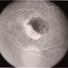
 Histoplasmosis and Subfoveal Neovascular Membrane
Mar 27 2019 by Gary R. Cook, MD, FACS
Mid-phase (20.4 seconds) fluorescein angiogram image of the right eye of 59-year-old white male with ocular histoplasmosis and a well-defined subfoveal CNVM OD; V.A.= 20/80+2
Imaging device: Topcon VT-50
Condition/keywords: FA mid phase, fluorescein angiogram (FA), ocular histoplasmosis syndrome (OHS), peripapillary atrophy, presumed ocular histoplasmosis syndrome (POHS), subfoveal choroidal neovascularization, subfoveal neovascular membrane
-
 Pathologic Myopia
Apr 2 2019 by Gary R. Cook, MD, FACS
Mid-phase fluorescein angiogram image of the right eye of 51-year-old white male with -25D myopia; V.A. = 20/70-1
Imaging device: Topcon VT-50
Condition/keywords: FA mid phase, fluorescein angiogram (FA), high myopia, pathologic myopia
-
 Pathologic Myopia
Apr 2 2019 by Gary R. Cook, MD, FACS
Mid-phase fluorescein angiogram image of the left eye of a 51-year-old white male with -25D myopia; V.A. = 20/40
Imaging device: Topcon VT-50
Condition/keywords: FA mid phase, fluorescein angiogram (FA), high myopia, pathologic myopia
-
---thumb.jpg/image-square;max$300,300.ImageHandler) Pattern Dystrophy
Pattern Dystrophy
Aug 9 2013 by From the Collections of Thomas M. Aaberg, MD and Thomas M. Aaberg Jr., MD
Mid phase FA of patient with pattern dystrophy.
Condition/keywords: FA mid phase, pattern macular dystrophy
-
 Posterior Scleritis
Jun 6 2019 by Gary R. Cook, MD, FACS
Mid-phase fluorescein angiogram image of acute posterior scleritis lesion beneath the superotemporal arcade OD; V.A. = 20/40-2
Imaging device: Topcon VT-50
Condition/keywords: FA mid phase, fluorescein angiogram (FA), posterior scleritis
-
 Posterior Uveitis
Apr 8 2019 by Gary R. Cook, MD, FACS
Mid-phase (64 seconds) fluorescein angiogram image showing mild leakage and early staining of the yellow-white spots in the temporal macula of the right eye; V.A. = 20/30.
Imaging device: Topcon VT-50
Condition/keywords: FA mid phase, fluorescein angiogram (FA), posterior uveitis
-
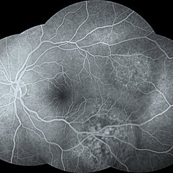
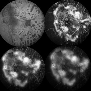 Proliferative Diabetic Retinopathy
May 28 2016 by Olivia Rainey
Fluorescein angiogram series of an 30-year-old male with proliferative diabetic retinopathy affecting his right eye. The patient presented with worsening neovascularization and scar tissue contracting in macula in the right eye. He experienced a decline in vision secondary to macula ischemia. Patient was seeing 20/400 and with PH 20/200 in the right eye and HM in the left eye.
Photographer: Olivia Rainey
Imaging device: Heidelberg Spectralis
Condition/keywords: diabetes, FA early phase, FA late phase, FA mid phase, fluorescein leakage, fundus autofluorescence (FAF), neovascularization (NV), proliferative diabetic retinopathy (PDR)
-
 Retinal Angiomatous Proliferation
Sep 10 2018 by Gabriela Lopezcarasa Hernandez, MD
75-year-old patient with decrease in visual acuity right eye with metamorphopsia, in the FA and ICG we can see a RAP lesion.
Photographer: Azucena Rios
Imaging device: Heidelberg Spectralis
Condition/keywords: FA mid phase, indocyanine green (ICG) angiography, RAP lesion, retinal angiomatous proliferation (RAP)
-
 RPE RIP
Jun 6 2019 by Gary R. Cook, MD, FACS
Mid-phase fluorescein angiogram of an elderly white female with exudative AMD and a RPE rip OS; V.A. = 20/80
Condition/keywords: FA mid phase, fluorescein angiogram (FA), retinal pigment epithelium
-
 Syphilitic Chorioretinitis
Apr 8 2019 by Gary R. Cook, MD, FACS
Mid-phase (48 seconds) fluorescein angiogram image of the right eye of a 68-year-old female with diffuse salt-and-pepper-type chorioretinitis secondary to syphilis; V.A. = 20/25
Imaging device: Topcon VT-50
Condition/keywords: chorioretinitis, FA mid phase, fluorescein angiogram (FA), pseudo retinitis pigmentosa, syphilis
-
 Vogt-Koyanagi-Harada with Multiple PEDs
Oct 10 2012 by Jeffrey G. Gross, MD, FASRS
VKH with multiple PEDs, FA mid phase.
Condition/keywords: FA mid phase, pigment epithelial detachment (PED)
 Loading…