Search results (48 results)
-
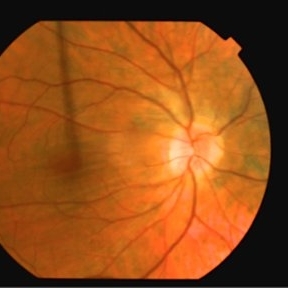
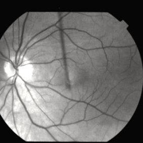
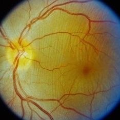
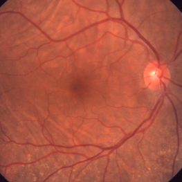 Bilateral Idiopathic Choroidal Folds
Jan 11 2013 by Gerardo Garcia-Aguirre, MD
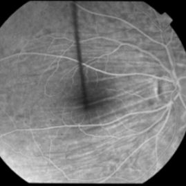
Fundus photograph of the right eye showing choroidal folds.
Imaging device: Zeiss FF4
Condition/keywords: choroidal folds
-
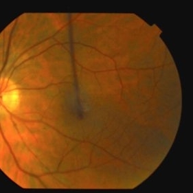
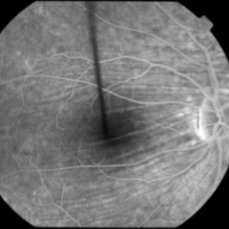
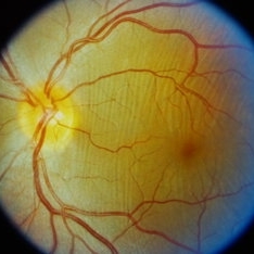
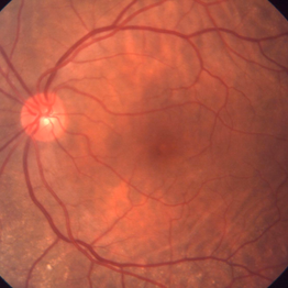 Bilateral idiopathic choroidal folds
Jan 11 2013 by Gerardo Garcia-Aguirre, MD
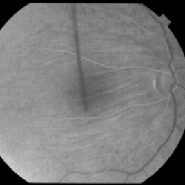
Fundus photograph showing choroidal folds.
Imaging device: Zeiss ff4
Condition/keywords: choroidal folds
-
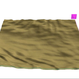 Bilateral Idiopathic Choroidal Folds
Jan 11 2013 by Gerardo Garcia-Aguirre, MD
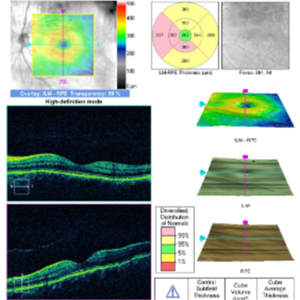
3D reconstruction of the RPE showing choroidal folds.
Photographer: Gerardo Garcia-Aguirre, MD
Imaging device: Zeiss Cirrus HD OCT
Condition/keywords: choroidal folds
-
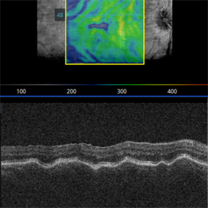
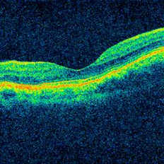 Bilateral Idiopathic Choroidal Folds - OCT
Jan 11 2013 by Gerardo Garcia-Aguirre, MD
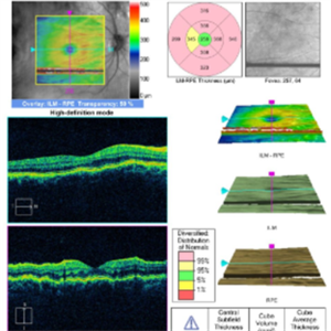
OCT of the macula showing choroidal folds.
Photographer: Gerardo Garcia-Aguirre, MD
Imaging device: Zeiss Cirrus HD OCT
Condition/keywords: choroidal folds
-
 Choroidal Folds
-
 Choroidal Folds
-
 Choroidal Folds
Nov 28 2014 by Thomas A. Ciulla, MD, MBA, FASRS
This 53-year-old man was noted to have choroidal folds right greater than left. The visual acuity was normal at 20/15. The choroidal folds are visible on OCT, especially on the vertical cuts that image across the horizontal folds. Angiography revealed staining of the folds without CNVM, choroidal mass, or optic nerve edema.
Photographer: Charlotte Harris
Condition/keywords: bilateral chorioretinal folds, choroidal folds
-
 Choroidal Folds
Nov 28 2014 by Thomas A. Ciulla, MD, MBA, FASRS
This 53-year-old man was noted to have choroidal folds right greater than left. The visual acuity was normal at 20/15. The choroidal folds are visible on OCT, especially on the vertical cuts that image across the horizontal folds. Angiography revealed staining of the folds without CNVM, choroidal mass, or optic nerve edema.
Condition/keywords: bilateral chorioretinal folds, choroidal folds
-
 Choroidal Folds
Nov 28 2014 by Thomas A. Ciulla, MD, MBA, FASRS
This 53 -year-old man was noted to have choroidal folds right greater than left. The visual acuity was normal at 20/15. The choroidal folds are visible on OCT, especially on the vertical cuts that image across the horizontal folds. Angiography revealed staining of the folds without CNVM, choroidal mass, or optic nerve edema.
Condition/keywords: bilateral chorioretinal folds, choroidal folds
-
 Choroidal Folds
Feb 21 2022 by Maxwell J Wingelaar, MD
61-year-old male with choroidal folds
Photographer: Jarrod Wehmeier
Condition/keywords: choroidal folds
-
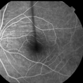
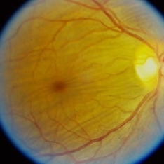
 Choroidal folds
Sep 21 2023 by Ben Serar
Fundus photograph of LE showing choroidal folds at the macula.
Condition/keywords: choroidal folds
-
 Choroidal folds
Sep 14 2023 by Ben Serar
Fundus photograph of the LE showing choroidal folds.
Condition/keywords: choroidal folds
-
 Choroidal folds
Sep 14 2023 by Ben Serar
Fundus photograph of RE showing choroidal folds at the posterior pole .
Condition/keywords: choroidal folds
-
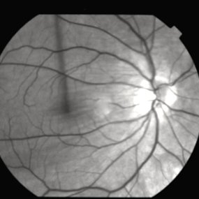
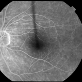
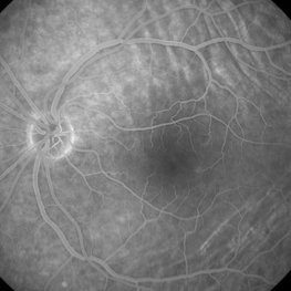 Choroidal Folds - Fluorescein Angiogram
Jan 11 2013 by Gerardo Garcia-Aguirre, MD
Fluorescein angiogram.
Photographer: Gerardo Garcia-Aguirre, MD
Imaging device: Zeiss FF4
Condition/keywords: choroidal folds
-
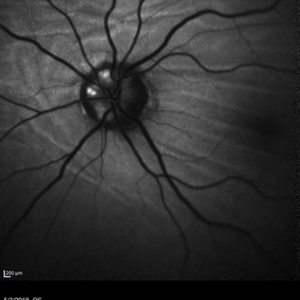 Choroidal Folds and Optic Disc Drusen
Aug 1 2018 by Emily Cooper
Fundus autofluorescence photo of a 62-year-old man who presented for evaluation of choroidal folds and optic disc drusen. He is currently following up with neuro-ophthalmology and has suspected intracranial hypertension.
Photographer: Emily Cooper, Retina Specialists of Michigan
Condition/keywords: choroidal folds, drusen of optic disc
-
 Choroidal folds due to hypotony
Jan 11 2013 by Alex P. Hunyor, MD
Choroidal folds due to hypotony
Condition/keywords: choroidal folds, hypotonous retinopathy
-
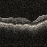 Choroidal folds i/c/o hypotony
Nov 23 2023 by Anand Temkar
OCT showing choroidal folds in a follow up case of filtration surgery with mitomycin c and anterior vitrectomy elsewhere.
Photographer: Dr.Anand Temkar- Retina Foundation, Ahmedabad
Imaging device: Mirante
Condition/keywords: choroidal folds, hypotony, OCT
 Loading…