Search results (5 results)
-
 Alport's Syndrome
Aug 29 2018 by Abhishek Das, MBBS, MS
OCT of a 54-year-old woman diagnosed to have Alport's syndrome. OCT shows temporal thinning of retina with nasal retina preserved.
Photographer: Abhishek Das, The Eye Foundation,Coimbatore,India
Imaging device: Optovue
Condition/keywords: Alports disease
-
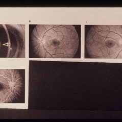 Alports Disease
Jul 29 2013 by H. Michael Lambert, MD
Alports disease, macular diseases not associated with FA changes. Spotty window defects with mid peripheral lesions. Disease may represent ABNL in basement membranes. Basal laminar drusen.
Condition/keywords: Alports disease
-
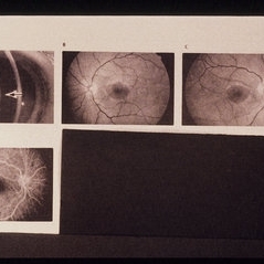 Alports Disease
Jul 29 2013 by H. Michael Lambert, MD
Alports disease, macular diseases not associated with FA changes. Spotty window defects with mid peripheral lesions. Disease may represent ABNL in basement membranes. Basal laminar drusen.
Condition/keywords: Alports disease
-
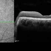 Plateau Fovea with Inner Retinal Thinning
May 27 2020 by Olivia Rainey
Optical coherence tomography of the left eye of a 20-year-old male with Alport Syndrome. The patient did not present with any ocular or visual symptoms, yet the distinct "plateau contour" of his fovea was noted on OCT during his visit. The patient presented with 20/25 vision at the time of his visit. There was myelinated nerve fiber layer noted in both eyes, but these features had remained stable from his appointment three years prior. The physician noted that myelinated nerve fiber was a congenital change, and had not affected his vision or health of the eye, nor is a feature of Alport Syndrome.
Photographer: Olivia Rainey, OCT-C, COA
Imaging device: Heidelberg Spectralis
Condition/keywords: Alports disease, Heidelburg Spectralis, inner retinal thinning, left eye, optical coherence tomography (OCT), plateau fovea
-
 Whole Eye OCT
Jan 4 2019 by Netan Choudhry, MD, FRCS(C) FASRS
Swept-Source OCT montage of a 45-year-old male with Alports disease and posterior subcapsular cataract.
Photographer: John Golding BA, Vitreous Retina Macula Specialists of Toronto
Imaging device: Topcon DRI Triton
Condition/keywords: Alports disease, optical coherence tomography (OCT), swept source
 Loading…