Search results (45 results)
-
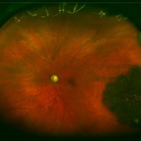
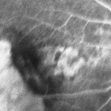
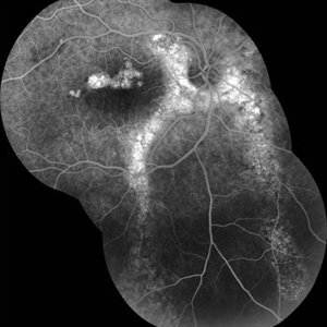
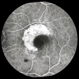 ARMD with RPE Rip
Oct 12 2012 by Jeffrey G. Gross, MD, FASRS
ARMD with RPE rip, FA, showing window defect and blockage from retracted RPE layer.
Condition/keywords: retinal pigment epithelium, retinal pigment epithelium (RPE) tear, retracted retinal pigment epithelium (RPE) layer
-
 Retinitis pigmentosa AD Slide 4
Oct 22 2012 by Ronald C. Gentile, MD
Early fluorescein angiography revealed early hyper-fluorescence surrounding the fovea consistent with retinal pigment epithelial (RPE) depigmentation. This accentuated the contrast between the normal blockage (hypo-fluorescence) of the macular luteal pigment from the surrounding RPE window defect (hyper-fluorescence).
Photographer: The New York Eye & Ear Infirmary Department of Medical Imaging
Condition/keywords: retinitis pigmentosa
-
 Pattern Macular Dystrophy
Oct 16 2012 by Ratimir Lazic, MD, PhD
FAG image of a 76-year-old female. In early venous phase window defect in fovea can be seen.
Photographer: Marko Lukic, MD
Imaging device: Zeis Visucam Lite 2
Condition/keywords: fundus photograph, retinal pigment epithelium (RPE) defect
-
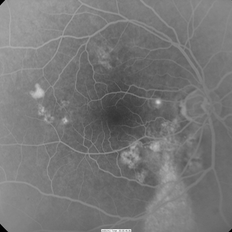
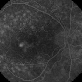 CNV due to AMPPE
Oct 16 2012 by Ratimir Lazic, MD, PhD
FAG of 58-year-old male. In late venous phase hyperflorescence of white dots (caused by window defect) can be seen. Intensive leakage of dye in juxtafoveolar region.
Photographer: Marko Lukic, MD
Imaging device: Zeis Visucam Lite 2
Condition/keywords: acute posterior multifocal placoid pigment epitheliopathy (APMPPE), choroidal neovascularization (CNV)
-
 Adenocarcinoma Arising from CHRPE
Sep 17 2015 by Marc C. Peden, MD
49-year-old female referred for presumed ocular melanoma. On examination was noted to have darkly pigmented lesion in the temporal retina of left eye. Lesion had characteristic scalloped edges with central lacunae, however, on ultrasonography was noted to have 1.8mm of elevation with high internal reflectivity. IVFA shows absence of dual circulation with areas of window defect. Findings were consistent with those described by Shields et al., in their April 2001 article in Archives of Ophthalmology.
Photographer: Janet Traynom
Imaging device: Optos P200MA
Condition/keywords: adenocarcinoma arising from CHRPE
-
---thumb.jpg/image-square;max$300,300.ImageHandler) Pattern Dystrophy
Pattern Dystrophy
Aug 7 2013 by From the Collections of Thomas M. Aaberg, MD and Thomas M. Aaberg Jr., MD
Fluoresceing angiography in the early laminar phase shows blocked fluorescence of the pigmented lesion and surrounding hyperfluorescense due to window defect.
Condition/keywords: butterfly dystrophy, pattern macular dystrophy
-
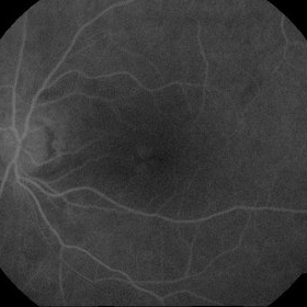
 Central areolar atrophy
May 2 2013 by Henry J. Kaplan, MD
Fluorescein angiogram demonstrates early hyperfluorescence due to window defect; the patient is young with GA like lesion and vision of 20/200 which is compatible with central areolar atrophy#1
Condition/keywords: central areolar choroidal dystrophy (CACD)
-
 Chronic Central Serous Chorioretinopathy
Oct 31 2012 by Lihteh Wu, MD
FA frame showing a hyperfluorescent window defect in a gutter pattern extending down from the posterior.
Condition/keywords: central serous chorioretinopathy (CSCR)
-
 CNV due to AMPPE
Oct 16 2012 by Ratimir Lazic, MD, PhD
FAG of 58-year-old male. In early venous phase hyperflorescence of white dots (caused by window defect) can be seen. Leakage of dye in juxtafoveolar region.
Photographer: Marko Lukic, MD
Imaging device: Zeis Visucam Lite 2
Condition/keywords: acute posterior multifocal placoid pigment epitheliopathy (APMPPE), choroidal neovascularization (CNV)
-
---thumb.jpg/image-square;max$300,300.ImageHandler) Age Related Macular Degeneration - Geographic Atrophy
Age Related Macular Degeneration - Geographic Atrophy
May 3 2013 by Suber S. Huang, MD, MBA, FASRS
Geographic Atrophy.
Imaging device: Retina Diseases Imaging Reading Center
Condition/keywords: advanced geographic atrophy, atrophic scar, atrophic spot, geographic atrophy, macula lesion, pigment epithelial atrophy, red-free, window defect
-
---thumb.jpg/image-square;max$300,300.ImageHandler) Age Related Macular Degeneration
Age Related Macular Degeneration
May 3 2013 by Suber S. Huang, MD, MBA, FASRS
Age related macular degeneration.
Condition/keywords: advanced geographic atrophy, atrophic scar, atrophic spot, geographic atrophy, macula lesion, pigment epithelial atrophy, red-free, window defect
-
 Toxocara Granuloma
Jun 4 2014 by Henry J. Kaplan, MD
Arteriovenous phase angiogram of the same patient shows staining of the granuloma and stippling hyperfluorescence around the lesion secondary to RPE window defect. #3
Condition/keywords: toxocara granuloma, toxocariasis
-
---thumb.jpg/image-square;max$300,300.ImageHandler) Cone Dystrophy
Cone Dystrophy
Feb 20 2013 by From the Collections of Thomas M. Aaberg, MD and Thomas M. Aaberg Jr., MD
FA of OS showing window defects in a circular pattern at the macula.
Condition/keywords: bull's eye maculopathy, cone dystrophy
-
---thumb.jpg/image-square;max$300,300.ImageHandler) RPE Hypertrophy
RPE Hypertrophy
Aug 8 2013 by From the Collections of Thomas M. Aaberg, MD and Thomas M. Aaberg Jr., MD
FA of the same patient. Shows typical window defects in the lacunar area due to chorioretinal atrophy #2.
Condition/keywords: retinal pigment epithelium (RPE) hypertrophy
-
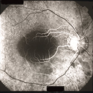
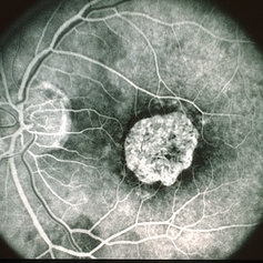
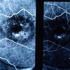
 RPE Tear: Fluorescein Angiography
May 2 2015 by Thomas A. Ciulla, MD, MBA, FASRS
Mid Phase Fluorescein Angiogram: The scrolled and redundant RPE just temporal to the fovea blocks underlying choroidal fluorescence. The absent RPE, more temporally, results in a window defect with intense hyperfluorescence.
Photographer: Stuart Alfred
Condition/keywords: choroidal neovascular membrane (CNVM), retinal pigment epithelium (RPE) tear, wet age-related macular degeneration (wet AMD)
-
 Chronic Central Serous Chorioretinopathy
Oct 31 2012 by Lihteh Wu, MD
FA frame showing a hyperfluorescent window defect in a gutter pattern. There is also a hot spot in the nasal macula.
-
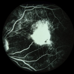
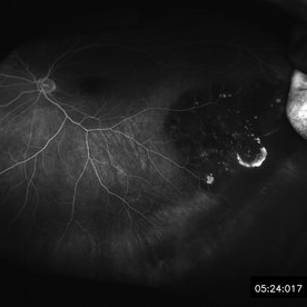
 PPCNVM and Peripheral Drusen Seen on Optos FA
Apr 22 2020 by John S. King, MD
72-year-old white male c/o of distortion OS for about 2 months. 20/100 OS, normotensive, small grey-green subretinal area just temporal to the optic disc. FA shows leakage c/w a ppcnvm; there is some SR and IR leakage as well as staining of peripheral drusen and some window defects from cobblestone. Avastin was adminstered.
Photographer: Asli Ahmed
Imaging device: CA
Condition/keywords: drusen, peripapillary choroidal neovascularization (PPCNVM)
-
 Adenocarcinoma Arising from CHRPE
Sep 17 2015 by Marc C. Peden, MD
49-year-old female referred for presumed ocular melanoma. On examination was noted to have darkly pigmented lesion in the temporal retina of left eye. Lesion had characteristic scalloped edges with central lacunae, however, on ultrasonography was noted to have 1.8mm of elevation with high internal reflectivity. IVFA shows absence of dual circulation with areas of window defect. Findings were consistent with those described by Shields et al., in their April 2001 article in Archives of Ophthalmology.
Photographer: Janet Traynom COT
Imaging device: Optos P200MA
Condition/keywords: adenocarcinoma arising from CHRPE
-
 Chronic CSCR - RPE Tracts
May 4 2014 by Neha Goel, MS DNB FRCS (Glasg)
FFA of a patient with chronic CSCR showing RPE window defects at the macula and RPE tracts running inferiorly from the peripapillary region.
Photographer: Neha Goel
Imaging device: Zeiss Visucam
Condition/keywords: chronic central serous chorioretinopathy (CSCR), retinal pigment epithelium
-
 Rubella Retinopathy
May 2 2013 by Henry J. Kaplan, MD
Fluorescein angiography of the same patient with rubella retinopathy demonstrates RPE window defects as hyperfluorescent areas; #3.
Condition/keywords: rubella retinopathy
-
 Bull's Eye
May 2 2013 by Henry J. Kaplan, MD
Fluorescein angiography demonstrates RPE window defects and hyperfluorescence in bull's eye maculopathy; #2.
Condition/keywords: bull's eye maculopathy
-
 Polypoidal Chroidal Vasculopathy
Sep 21 2018 by Dhaivat Shah
A 40-year-old female presented with sudden onset decreased vision in right eye. BCVA: CF 1 mt. Fundus showed massive subretinal exudation with haemorrhage. EDI OCT showed notched PEDs with shallow SRF and exudation with back-shadowing. FFA shows leak with window defects. ICG shows hotspot in late phase. Polypoidal choroidal vasculopathy (PCV) is a retinal disorder characterized by the presence of aneurysmal polypoidal lesions in the choroidal vasculature, resulting in damage to the overlying retina and loss of retinal pigment epithelium. The aneurysmal dilatations, also known as polyps, may be found subfoveal, juxtafoveal, extrafoveal, peripapillary or even peripheral regions. The polypoidal lesions are best detected on indocyanine green angiography as hotspots in late phase. The presence of choroidal polyps can lead to recurrent episodes of exudative retinal detachment, serous or hemorrhagic pigment epithelial detachment, subretinal hemorrhage and exudation. Treatment is available in form of laser/PDT along with Anti VEGF injection.
Photographer: Miss Moupiya Das
Condition/keywords: polypoidal choroidal vasculopathy (PCV)
-
 Alports Disease
Jul 29 2013 by H. Michael Lambert, MD
Alports disease, macular diseases not associated with FA changes. Spotty window defects with mid peripheral lesions. Disease may represent ABNL in basement membranes. Basal laminar drusen.
Condition/keywords: Alports disease
-
 Torpedo Maculopathy
Jan 20 2020 by Pierre-Henry Gabrielle, MD
Coupled OCT B-scan and fluorescein angiogram of an asymptomatic 12-year-old girl with torpedo maculopathy of the left eye. One can report complete RPE atrophy at lesion site with window defect on FA and choroidal cavitation on OCT.
Photographer: Pierre-Henry Gabrielle, Ophthalmology department, Dijon University Hospital, France
Imaging device: Heidelberg Spectralis
Condition/keywords: fluorescein angiogram (FA), optical coherence tomography (OCT), torpedo maculopathy
-
 Macular Degeneration with Extensive Geographic Atrophy
Jan 26 2022 by Olivia Rainey
Heidelberg Spectralis fluorescein angiography of a 94-year-old woman with Macular Degeneration affecting both eyes. The FA reveals transmission defects consistent with RPE changes and geographic atrophy of RPE of both eyes, as well as window defects consistent with peripheral scarring in the right eye. The patient's vision was Dcc20/70 in both eyes at the visit the images were taken.
Photographer: Olivia Rainey, OCT-C, COA
Imaging device: Heidelberg Spectralis
Condition/keywords: 30-degrees, choroidal neovascularization (CNV), dry age-related macular degeneration (dry AMD), early phase, fluorescein angiogram (FA), geographic atrophy, heidelberg spectralis, macular degeneration, neovascular age-related macular degeneration (AMD)
 Loading…