Search results (15 results)
-
---thumb.jpg/image-square;max$300,300.ImageHandler) Asteroids-B Scan
Asteroids-B Scan
Apr 18 2014 by Susanna S. Park, MD, PhD
B-scan ultrasound image of the left eye of a 95-year-old Hispanic diabetic man with dense media opacity from asteroids hyalosis. Visual acuity is 20/60.
Photographer: Ellen Redenbo, UC Davis Eye Center
Condition/keywords: asteroid hyalosis, B scan ultrasound, vitreous opacity
-
 Remnant of Hyaloidal Artery
Feb 5 2014 by Gerardo Garcia-Aguirre, MD
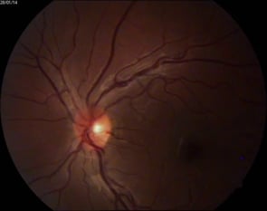
Fundus photograph of the left eye of a 14-year-old asymptomatic female. The photograph is focused on the retina, and a prepapillary vitreous opacity is observed (white arrows). The opacity is attached to the origin of the retinal vessels in the optic nerve head.
Photographer: Gerardo Garcia-Aguirre, MD
Condition/keywords: persistence of the hyaloid artery
-
 Remnant of Hyaloidal Artery
Feb 5 2014 by Gerardo Garcia-Aguirre, MD
Fundus photograph of the left eye of a 14-year-old asymptomatic female. The photograph is focused on the posterior vitreous where a prepapillary vitreous opacity is observed (white arrows). The opacity is attached to the origin of the retinal vessels in the optic nerve head.
Photographer: Gerardo Garcia-Aguirre, MD
Condition/keywords: persistence of the hyaloid artery
-
 Remnant of Hyaloidal Artery
Feb 5 2014 by Gerardo Garcia-Aguirre, MD
Video of the fundus of the left eye of a 14-year-old asymptomatic female, where a prepapillary vitreous opacity is observed. The opacity is attached to the origin of the retinal vessels in the optic nerve head, and is considered to be a remnant of the hyaloidal artery.
Photographer: Gerardo Garcia-Aguirre, MD
Condition/keywords: persistence of the hyaloid artery
-
 Remnant of Hyaloidal Artery
Feb 5 2014 by Gerardo Garcia-Aguirre, MD
Fundus photograph of the left eye of a 14-year-old asymptomatic female. The photograph is focused on the posterior vitreous where a prepapillary vitreous opacity is observed (see next picture where opacity is marked by arrows). The opacity is attached to the origin of the retinal vessels in the optic nerve head.
Photographer: Gerardo Garcia-Aguirre, MD
Condition/keywords: persistence of the hyaloid artery
-
 Vitreous Amyloidosis Slit Lamp Photo
Oct 23 2019 by Alexander D Port, MD
Slit lamp photograph preoperatively demonstrating dense symptomatic vitreous opacity in the setting of amyloidosis. The patient elected to undergo pars plana vitrectomy.
Condition/keywords: slit lamp photo, vitreous amyloidosis
-
 Remnant of Hyaloidal Artery
Feb 5 2014 by Gerardo Garcia-Aguirre, MD
Fundus photograph of the left eye of a 14-year-old asymptomatic female. The photograph is focused on the retina, and a prepapillary vitreous opacity is observed (white arrows). The opacity is attached to the origin of the retinal vessels in the optic nerve head.
Photographer: Gerardo Garcia-Aguirre, MD
Condition/keywords: persistence of the hyaloid artery
-
 Vitreous Amyloidosis Slit Lamp Photo
Oct 23 2019 by Alexander D Port, MD
Slit lamp photograph preoperatively demonstrating dense symptomatic vitreous opacity in the setting of amyloidosis. The patient elected to undergo pars plana vitrectomy.
Condition/keywords: slit lamp photo, vitreous amyloidosis
-
 Chronical Submacular Hemorrhage in the Setting of Neovascular AMD
Mar 23 2015 by Rita Couceiro, MD, MS
An 80-year-old male, with a history of hypertension and high cholesterol, complained of acute and painless vision loss in his left eye (OS) in the previous 5 months. On observation best corrected visual acuity in OS was hand motion. A dense vitreous opacity in OS precluded fundus examination. Ocular ultrasound revealed vitreous hemorrhage and thickening of the macular area. The patient was submitted to pars plana vitrectomy, which disclosed a large submacular hemorrhage with chronical features and disciform scarring in the setting of neovascular AMD.
Imaging device: Intraoperative fundus photograph
Condition/keywords: neovascular age-related macular degeneration (AMD), submacular hemorrhage, wet age-related macular degeneration (wet AMD)
-
---thumb.jpg/image-square;max$300,300.ImageHandler) Fundus Photography and Fluorescein Angiography of Candida Endopthalmitis.
Fundus Photography and Fluorescein Angiography of Candida Endopthalmitis.
Dec 24 2013 by Dong Yoon Kim, MD
71-year-old woman visited our clinic for vitreous opacity of her right eyes. 2 weeks ago, she underwent ocular trauma for her right eyes. She had no systemic disease. Vitrectomy and vitreous culture was performed for the vitreous opacity. Candida albicans was identified from the vitreous culture.
Condition/keywords: candida endophthalmitis, fundus photograph
-
 Vitreous Amyloidosis Pre-Op
Oct 23 2019 by Alexander D Port, MD
Optos ultra wide field image preoperatively demonstrating dense symptomatic vitreous opacity in the setting of amyloidosis. The patient elected to undergo pars plana vitrectomy.
Condition/keywords: vitreous amyloidosis
-
 Vitreous Amyloidosis Postop
Oct 23 2019 by Alexander D Port, MD
Optos ultra wide field image postoperatively demonstrating clearance of vitreous opacity s/p pars plana vitrectomy.
Condition/keywords: vitreous amyloidosis
-
 Slide 2-36
Feb 19 2019 by Lancaster Course in Ophthalmology
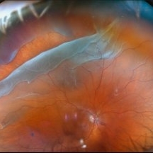
Involvement of the pars plana region in a 55-year-old woman with sarcoid. "Snow bank" exudate over the ciliary body and large vitreous opacities resemble those in the clinical entity "pars planitis."
Condition/keywords: ciliary, pars plana, pars planitis, sarcoid, vitreous opacity
-
 Asteroid Hyalosis
Dec 16 2018 by Hashim Ali Khan, OD, FAAO
Slit-lamp photographs of a 50-year-old man with asteroid hyalosis.
Condition/keywords: asteroid hyalosis, vitreous opacity
-
 Retinal Detachment with Giant Retinal Tear
May 14 2025 by Kimberly Wakester
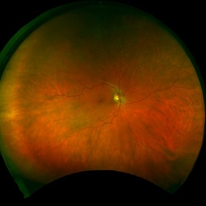
Optomap RGB of an 66-year-old man with a retinal detachment with a giant retinal tear in the right eye. Surgery was recommended. Patient is to continue follow up care post operatively. Also noted in the image is a vitreous opacity that was caught at the right moment and appears to look like a smiley face.
Photographer: Kimberly Wakester, COA, OCT-C
Imaging device: Optos California
Condition/keywords: giant retinal tear, RD
 Loading…