Search results (9 results)
-
 Syphilis Neuroretinopathy
Apr 2 2018 by JEFFERSON R SOUSA, Tecg.º (Biomedical Systems Technology)
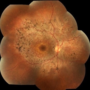
Female patient, 21-years-old, with complaint of low vision in the right eye for 3 years. According to information from the patient's history, at the time she noticed the low vision, it also coincided with a picture of a strong urinary infection as well as episodes of constant tonsillitis. Yes, the patient did not seek medical attention and self-medicated with antibiotics. In ophthalmologic evaluation, as well as examinations of color retinography and ocular fundus autofluorescence, important pigmentary alterations were observed following vascular arches with pigment mobilization in osteoclasts (aspect of a unilateral pigmentary retinitis secondary to the inflammatory process). Which suggested inflammatory process sequelae. Through the laboratory tests, he had positive (+) confirmation for SYPHILIS NEURORETINOPATHY .
Photographer: JEFFERSON R SOUSA - Study Center and Ophthalmological Research Dr. Andre M V Gomes, Institute Dr. Suel Abujamra São Paulo-Brazil
Imaging device: Fundus camera Topcon TRC-50 DX, Imaginet 5.0, angle de 50 graus. Flash 36 / Mosaic with 10 images.
Condition/keywords: neurosyphilitic optic atrophy, retinitis pigmentosa, syphilis, syphilis neuroretinopathy
-
 Syphilis Neuroretinopathy FA
Sep 25 2013 by Alexandre Durao Alves Pereira, MD
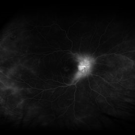
Syphilis neuroretinopathy, late phase FA.
Photographer: Alexandre Pereira
Condition/keywords: FA late phase
-
 Syphilis Neuroretinopathy (Color Photo)
Sep 25 2013 by Alexandre Durao Alves Pereira, MD
Syphilis neuroretinopathy, late phase FA, (color photograph).
Photographer: Alexandre Pereira
Condition/keywords: color photo, late phase, syphilis neuroretinopathy
-
 Syphilis Neuroretinopathy
Sep 25 2013 by Alexandre Durao Alves Pereira, MD
Syphilis neuroretinopathy late phase FA.
Photographer: Alexandre Pereira
Condition/keywords: late phase
-
 Syphilis Neuroretinopathy (Red Free Photo)
Sep 25 2013 by Alexandre Durao Alves Pereira, MD
Syphilis neuroretinopathy, late phase FA, (red free photograph).
Photographer: Alexandre Pereira
Condition/keywords: late phase, red-free
-
 Syphilis Neuroretinopathy (Late Phase FA)
Sep 25 2013 by Alexandre Durao Alves Pereira, MD
Syphilis neuroretinopathy, (late phase FA).
Photographer: Alexandre Pereira
Condition/keywords: late phase
-
 Syphilis Neuroretinopathy
Apr 2 2018 by JEFFERSON R SOUSA, Tecg.º (Biomedical Systems Technology)
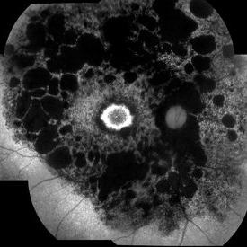
Female patient, 21-years-old, with complaint of low vision in the right eye for 3 years. According to information from the patient's history, at the time she noticed the low vision, it also coincided with a picture of a strong urinary infection as well as episodes of constant tonsillitis. Yes, the patient did not seek medical attention and self-medicated with antibiotics. In ophthalmologic evaluation, as well as examinations of color retinography and ocular fundus autofluorescence, important pigmentary alterations were observed following vascular arches with pigment mobilization in osteoclasts (aspect of a unilateral pigmentary retinitis secondary to the inflammatory process). Which suggested inflammatory process sequelae. Through the laboratory tests, he had positive (+) confirmation for SYPHILIS NEURORETINOPATHY .
Photographer: JEFFERSON R SOUSA - Study Center and Ophthalmological Research Dr. Andre M V Gomes, Institute Dr. Suel Abujamra São Paulo-Brazil
Imaging device: Fundus camera Topcon TRC-50 DX, Imaginet 5.0, angle de 50 graus. Flash 100 / Mosaic with 10 images.
Condition/keywords: autofluorescence imaging, neurosyphilitic optic atrophy, retinitis pigmentosa, syphilis, syphilis neuroretinopathy
-
 Syphilis Neuroretinopathy (Late Phase FA)
Sep 25 2013 by Alexandre Durao Alves Pereira, MD
Syphilis neuroretinopathy, (late phase FA).
Photographer: Alexandre Pereira
Condition/keywords: late phase
-
 Syphilitic Uveitis
Apr 2 2020 by Olivia Rainey
Ultrawide-field fluorescein angiogram of a 42-year-old male with syphilitic uveitis affecting his right eye more than his left. Patient is HIV positive. He developed hearing loss and palm/leg/scalp rash prompting diagnosis of neurosyphilis, s/p IM and full IV course of 2.4 Mil PCN G, and finished this course 3/9/20. He admits to recent rectal bleeding with ongoing plan for colonoscopy 3/16/20. He has a history of extensive travel including London, Hong Kong, and Bangkok. His husband has also been treated with IV PCN G, however per chart review he has multiple sexual partners. Patient's vision was 20/20 in each eye.
Photographer: Olivia Rainey
Imaging device: Optos California
Condition/keywords: disc hyperfluorescence, FA late phase leakage, fluorescein angiogram (FA), fluorescein leakage, HIV, late phase, optic nerve edema, Optos, phelbitis, syphilis neuroretinopathy, ultra-wide field imaging, uveitis
 Loading…