Search results (74 results)
-
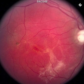
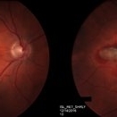
 Inferior Rhegmatogenous Retinal Detachment with Subretinal Fibrosis
Aug 23 2012 by Gabriela Lopezcarasa Hernandez, MD
Asymptomatic 25-year-old woman with high myopia.
Photographer: Gabriela Lopezcarasa Hernandez, Hospital Angeles Lomas
Imaging device: FF4
Condition/keywords: high myopia, subretinal fibrosis
-
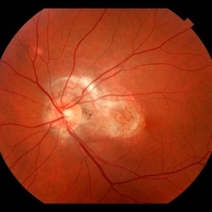
 Central Serous Chorioretinopathy (CSC)
Oct 16 2012 by S. Natarajan, MD, FASRS, FRCS (GLASGOW) , FICO, D.Sc, FELA
Middle-aged male came with small PED 4 months back; now this has progressed to a larger PED with SRF underneath the fovea.
Photographer: Prof. Dr. S. Natarajan
Condition/keywords: central serous chorioretinopathy (CSCR), central serous retinopathy (CSR), pigment epithelial detachment (PED), subretinal fibrosis
-
 Diffuse Choroidal Hemangioma
Nov 7 2012 by Rajiv Anand, MD, FRCS, FASRS
Fundus photo shows classic 'tomato-ketchup' red appearance of diffuse hemangioma. Due to chronic SRF , there is subretinal fibrosis.
Condition/keywords: subretinal fibrosis
-
 Angioid streaks - PXE
Jan 11 2013 by Alex P. Hunyor, MD
Pseudoxanthoma elasticum with angioid streaks, left eye - note subretinal fibrosis adjacent to disc.
Condition/keywords: angioid streaks, pseudoxanthoma elasticum (PXE)
-
 Subretinal Fibrosis (PPCNVM and POHS) OS
Sep 18 2019 by John S. King, MD
57-year-old white male with history of PPCNVM OS and POHS OU here for a routine visit. History of avastin in 2014, and stable since then. Va OS 20/20. PP scar with macular subretinal fibrosis. No heme or exudates. CR spot supero-nasally.
Photographer: Shelly Blair
Imaging device: Topcon 50
Condition/keywords: choroidal neovascular membrane (CNVM), ocular histoplasmosis syndrome (OHS), peripapillary choroidal neovascularization (PPCNVM), presumed ocular histoplasmosis syndrome (POHS)
-
 Angioid Streaks With CNV
Mar 11 2014 by Andrew M Hendrick, MD
Fundus photography of the right eye of a 50-year-old African American male with a remote history of minor trauma. Serial anti-VEGF injections failed to improve the subfoveal CNV and his condition is now being observed.
Photographer: Jannah Dobbs
Condition/keywords: angioid streaks, subretinal fibrosis
-
 Rhegmatogenous Retinal Detachment
Oct 11 2013 by Jason S. Calhoun
Patient in for a second opinion on RD, right eye. VA is NLP in the right eye. Fundus photography shows inferior retinal detachment with holes and subretinal fibrosis. No further surgery is suggested at this time.
Photographer: Jason S. Calhoun, Ophthalmic Photographer, Department of Ophthalmology, Mayo Clinic Jacksonville
Imaging device: TOPCON TRC 50-EX
-
 multifocal choroiditis
Feb 14 2013 by From the Collections of Thomas M. Aaberg, MD and Thomas M. Aaberg Jr., MD
color fundus photos showing healed chorioretinal scars, pigment deposition, and subretinal fibrosis consistent with regressed multifocal choroiditis
Condition/keywords: multifocal choroiditis, posterior segment inflammation, subretinal fibrosis, white dot syndrome
-
 Rhegmatogenous Retinal Detachment
Oct 11 2013 by Jason S. Calhoun
Patient in for second opinion on RD, right eye. VA is NLP in the right eye. Fundus photography shows inferior retinal detachment with holes and subretinal fibrosis. No further surgery is suggested at this time.
Photographer: Jason S. Calhoun, Ophthalmic Photographer, Department of Ophthalmology, Mayo Clinic Jacksonville
Imaging device: TOPCON TRC 50-EX
-
 Angioid streaks - PXE case 2
Jan 11 2013 by Alex P. Hunyor, MD
Pseudoxanthoma elasticum with angioid streaks, right eye - one year after initial image, showing extensive subretinal fibrosis in the macula.
Condition/keywords: angioid streaks, pseudoxanthoma elasticum (PXE)
-
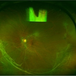
 Subretinal Fibrosis
Feb 20 2015 by H. Michael Lambert, MD
Posterior pole with possible subretinal fibrosis.
Condition/keywords: subretinal fibrosis
-
 Subretinal Thickening and Subretinal Hemorrhage – Stereo Color Fundus Photograph
Mar 9 2017 by James B. Soque, CRA, OCT-C, COA, FOPS
Color fundus stereo photograph of a 52-year-old white male with VA loss to 20/200 of unknown etiology. Dilated fundus examination of the right eye reveals a fibrotic scar with subretinal thickening and subretinal hemorrhage.
Photographer: James B Soque, CRA, OCT-C, COA
Imaging device: Topcon TRC 50 DX, MERGE Software
Condition/keywords: blood, color fundus photograph, color photo, stereo pair, subretinal blood, subretinal fibrosis, subretinal thickening
-
 Angioid Streaks
Mar 11 2014 by Andrew M Hendrick, MD
Fundus photography of the left eye of a 50-year-old African American male with a remote history of minor trauma in the contralateral eye.
Condition/keywords: angioid streaks, subretinal fibrosis
-
 Chorioretinal Scars with Subretinal Fibrosis and an old Retinal Detachment
May 3 2018 by Nichole Lewis
Chorioretinal scars with subretinal fibrosis and an old retinal detachment.
Photographer: Nichole Lewis
Condition/keywords: chorioretinal scar, chronic retinal detachment, subretinal fibrosis
-
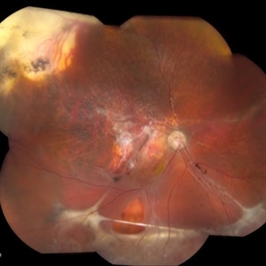
 Wagner Syndrome
Aug 1 2017 by Eitae Kim, MD
Ulltra wide field fundus photograph of 19-year-old male with Wagner syndrome which shows peripheral subretinal fibrosis and pigmentary degeneration.
Photographer: Eitae Kim, BOIM retinal center, Pureun eye hospital
Condition/keywords: subretinal fibrosis, ultra-wide field imaging, Wagner disease
-
 Peculiar Acute Subretinal Fibrosis
Feb 24 2015 by David Callanan, MD
Peculiar acute subretinal fibrosis.
Condition/keywords: subretinal fibrosis
-
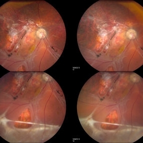
---thumb.jpg/image-square;max$300,300.ImageHandler) Bilateral Peripapillary Subretinal Fibrosis With Macula Choroidal Neovascularization
Bilateral Peripapillary Subretinal Fibrosis With Macula Choroidal Neovascularization
Dec 6 2013 by Maurice F. Rabb
Bilateral peripapillary subretinal fibrosis with macula choroidal neovascularization.
Condition/keywords: bilateral peripapillary subretinal fibrosis, macula choroidal neovascularization
-
 Peculiar Acute Subretinal Fibrosis
Feb 24 2015 by David Callanan, MD
Peculiar acute subretinal fibrosis.
Condition/keywords: subretinal fibrosis
-
---thumb.jpg/image-square;max$300,300.ImageHandler) Bilateral Peripapillary Subretinal Fibrosis With Macula Choroidal Neovascularization
Bilateral Peripapillary Subretinal Fibrosis With Macula Choroidal Neovascularization
Dec 6 2013 by Maurice F. Rabb
Bilateral peripapillary subretinal fibrosis with macula choroidal neovascularization.
Condition/keywords: bilateral peripapillary subretinal fibrosis, macula choroidal neovascularization
-
---thumb.jpg/image-square;max$300,300.ImageHandler) Bilateral Peripapillary Subretinal Fibrosis With Macula Choroidal Neovascularization
Bilateral Peripapillary Subretinal Fibrosis With Macula Choroidal Neovascularization
Dec 6 2013 by Maurice F. Rabb
Bilateral peripapillary subretinal fibrosis with macula choroidal neovascularization.
Condition/keywords: bilateral peripapillary subretinal fibrosis, macula choroidal neovascularization
 Loading…