Search results (8 results)
-
 Lattice Degeneration
Nov 9 2012 by Norman Byer
This is a more typical classical example of lattice degeneration in a 42-year-old woman in a photograph taken without scleral indentation. It shows much more marked vascular changes than the previous case. Note the tapering of the blood columns as the vessels approach the lesion and also the white sheathing of the vessel walls. Note also the continuity of the blood vessels on opposite sides of the lesion with the characteristic white lattice lines. More than 45 years ago Vogt pointed this out as a proof that these white lines were actually caused by changed blood vessels. Note also that this lesion shows a combination of several individual features of lattice degeneration. In addition to the white lines, there is a reddish crater-like area beneath the main horizontal white line. There is a prominent horizontal zone below this white line showing a snailtrack appearance. Also, there are two tiny atrophic retinal holes outside the photograph on the right end of this lesion. This eye contained five such retinal holes and they have all remained unchanged for more than 10 years of observation without treatment.
Condition/keywords: atrophic retinal hole, lattice degeneration, moderate snail track, tapering blood columns, white lattice lines, white sheath vessel
-
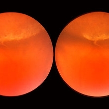
 Snail Track Peripheral Retinal Degeneration
Apr 29 2022 by Otakar Dušek, M.D. Ph.D.
Colour fundus photograph of 22-year-old woman with incidentally found snail track retinal degeneration in the superior temporal periphery of the retina of the right eye.
Photographer: Otakar Dušek, Charles University, Prague
Imaging device: Zeiss Clarus
Condition/keywords: peripheral retinal degeneration
-
 Lattice Degeneration
Nov 9 2012 by Norman Byer
This is lattice degeneration in a 10-year-old boy showing an almost pure snailtrack feature with only a hint of a reddish crater in the center. It has not changed over 10 years. The photograph was taken with scleral indentation.
Condition/keywords: lattice degeneration, reddish crater, scleral indentation, snail track
-
 Lattice Lesion
Nov 9 2012 by Norman Byer
This lattice lesion in a 30- year-old woman also shows combined features with a reddish crater above and a parallel snailtrack appearance just below it. Please note especially another interesting feature. From the left end of the lesion, there is a faint thin yellow line slanting down toward the right just below the shadow of the scleral indentation. This line identifies the dome of the pocket of liquified vitreous which is present over every lesion of lattice degeneration.
Condition/keywords: lattice degeneration, lattice lesion, liquefied vitreous, reddish crater, scleral indentation, snail track
-
 Lattice Lesion
Nov 9 2012 by Norman Byer
This lattice lesion in a 36-year-old woman has remained unchanged over a period of 13 years. It shows a moderate snailtrack feature with discrete yellow dots visible on the surface of the lesion and especially along the posterior border. One of these can be well seen just below the lesion superimposed over the dark shadow of the scleral indentation. The exact nature of these yellow dots is still not entirely clear.
Condition/keywords: lattice degeneration, moderate snail track, scleral indentation, yellow dots
-
 Lattice Combined with Tiny Round Hole
Nov 9 2012 by Norman Byer
This 45 year-old man shows the snail track form of lattice combined with a tiny round hole. There is a tiny subclinical retinal detachment confined to the lesion itself.
Condition/keywords: glial vitreous tuft, lattice degeneration, round hole, snail track
-
 Lattice Lesion
Nov 9 2012 by Norman Byer
This lattice lesion in a 36-year-old woman shows a snail track feature on the left combined with a reddish crater and retinal hole to the right. The hole has caused a small subclinical detachment. The next slide pair will show more of this lesion.
Condition/keywords: lattice lesion, reddish crater, retinal hole, snail track, subclinical detachment
-
 Lattice Lesion
Nov 9 2012 by Norman Byer
This lattice lesion in a 27-year-old woman shows an interesting change in the middle of the lesion. The predominant feature on the left side of the lesion is the snailtrack appearance while the right side of the lesion shows mainly a reddish crater. Note the many yellow dots above the surface of the retina which are actually located in the vitreous condensation which surrounds the pocket of liquified vitreous over the lesion.
Condition/keywords: lattice lesion, reddish crater, snail track, vitreous condensation, vitreous liquefaction
 Loading…