Search results (96 results)
-
---thumb.jpg/image-square;max$300,300.ImageHandler) Retinal Telangiectasia
Retinal Telangiectasia
Oct 18 2013 by Maurice F. Rabb
A 46 year old white male was seen with a history of retinal telangiectasia in the left eye.
Condition/keywords: retinal telangiectasia
-
 Myelinated retinal nerve fibers Slide 1
Oct 22 2012 by Ronald C. Gentile, MD
Myelinated retinal nerve fibers involving the inferior temporal arcade with associated retinal vascular abnormalities.
Photographer: The New York Eye & Ear Infirmary Department of Medical Imaging
Condition/keywords: myelinated nerve fibers, retinal telangiectasia
-
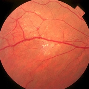
 Macular aneurysmal telangiectasia
Jan 11 2013 by Alex P. Hunyor, MD
Congenital retinal telangiectasia - Leber's miliary aneurysm end of the spectrum, with leak into macula
Condition/keywords: aneurysm, congenital retinal telangiectasis, retinal telangiectasia
-
 Myelinated retinal nerve fibers Slide 2
Oct 22 2012 by Ronald C. Gentile, MD
A magnified fundus photo of the myelinated retinal nerve fibers showing the retinal vascular abnormalities with multiple aneurysmal dilations.
Photographer: The New York Eye & Ear Infirmary Department of Medical Imaging
Condition/keywords: myelinated nerve fibers, retinal telangiectasia
-
---thumb.jpg/image-square;max$300,300.ImageHandler) Retinal Telangiectasia
Retinal Telangiectasia
Oct 18 2013 by Maurice F. Rabb
A 46 year old white male was seen with a history of retinal telangiectasia in the left eye.
Condition/keywords: retinal telangiectasia
-
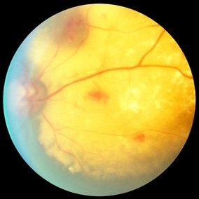
 Coats Disease
Oct 9 2012 by Alan D. Letson, MD
Coats Disease
Photographer: Beverly Radcliffe
Condition/keywords: retinal macroaneurysm, retinal telangiectasia
-
 Coats' Disease
Apr 27 2018 by Brenda Fallas
3-year-old boy with unilateral Coats' Disease fundus photo.
Photographer: Brenda Fallas, Bascom Palmer Eye Institute, Miami, FL
Imaging device: Retcam III 130 degree lens
Condition/keywords: Coats' disease, color fundus photograph, retinal telangiectasia
-
---thumb.jpg/image-square;max$300,300.ImageHandler) Type II Idiopathic Bilateral Juxtafoveolar Retinal Telangiectasis
Type II Idiopathic Bilateral Juxtafoveolar Retinal Telangiectasis
Oct 21 2013 by Maurice F. Rabb
DB2 was a 59 year old female first seen in November, 1982. She complained of declining vision in both eyes for four years. Initially she was in good general health, but ten years later she developed peripheral neuropathy and glucose intolerance. Diagnosis: Type II idiopathic bilateral juxtafoveolar retinal telangiectasis.
Condition/keywords: retinal telangiectasia
-
---thumb.jpg/image-square;max$300,300.ImageHandler) Coats Disease
Coats Disease
Oct 30 2012 by Lihteh Wu, MD
FA frame showing blocked fluorescence from the massive lipid exudation. There is also hyperfluorescence secondary to vascular leakage and hypofluorescence from the hyperplastic RPE. Superotemporal to the fovea there are areas of telangiectasia.
Condition/keywords: massive lipid exudation, retinal pigment epithelium, retinal telangiectasia
-
---thumb.jpg/image-square;max$300,300.ImageHandler) Type II Idiopathic Bilateral Juxtafoveolar Retinal Telangiectasis
Type II Idiopathic Bilateral Juxtafoveolar Retinal Telangiectasis
Oct 21 2013 by Maurice F. Rabb
DB2 was a 59 year old female first seen in November, 1982. She complained of declining vision in both eyes for four years. Initially she was in good general health, but ten years later she developed peripheral neuropathy and glucose intolerance. Diagnosis: Type II idiopathic bilateral juxtafoveolar retinal telangiectasis.
Condition/keywords: retinal telangiectasia
-
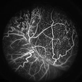 Coats' Disease FA
Apr 27 2018 by Brenda Fallas
3-year-old boy with unilateral Coats' Disease FA photo.
Photographer: Brenda Fallas, Bascom Palmer Eye Institute, Miami, FL
Imaging device: Retcam III 130 degree lens
Condition/keywords: Coats' disease, FA early phase, fluorescein angiogram (FA), retinal telangiectasia
-
 Coats Disease
Apr 16 2015 by Rita Couceiro, MD, MS
Fundus photograph and fluorescein angiography pictures of a 13-year-old girl with Coats Disease, showing abnormal telangiectatic vessels and intense exsudation in the inferior retinal periphery of the left eye.
Condition/keywords: Coats' disease, retinal telangiectasia
-
---thumb.jpg/image-square;max$300,300.ImageHandler) Type II Idiopathic Bilateral Juxtafoveolar Retinal Telangiectasis
Type II Idiopathic Bilateral Juxtafoveolar Retinal Telangiectasis
Oct 21 2013 by Maurice F. Rabb
DB2 was a 59 year old female first seen in November, 1982. She complained of declining vision in both eyes for four years. Initially she was in good general health, but ten years later she developed peripheral neuropathy and glucose intolerance. Diagnosis: Type II idiopathic bilateral juxtafoveolar retinal telangiectasis.
Condition/keywords: retinal telangiectasia
-
 Coats Disease Slide 1
Oct 22 2012 by Ronald C. Gentile, MD
A unilateral, sub-retinal, and yellowish exudative lesion with associated retinal telangiectasias involving the nasal retina. Refractile elements can be seen and represent cholesterol crystals.
Photographer: The New York Eye & Ear Infirmary Department of Medical Imaging
Condition/keywords: congenital retinal telangiectasis
-
---thumb.jpg/image-square;max$300,300.ImageHandler) Right Macular Lesion
Right Macular Lesion
Feb 3 2014 by Maurice F. Rabb
Male patient with lesion in the right macula. It appears he has a small area of epiretinal or intraretinal fibrotic or fibrotic tissue wiht nonperfusion encircled by telangienctasis.
Condition/keywords: macular lesion, retinal telangiectasia
-
 Congenital Juxtafoveal Telangiectasis
Mar 6 2014 by David Callanan, MD
17-year-old Hispanic male, congenital juxtafoveal telangiectasis.
Condition/keywords: juxtafoveal telangiectasis, retinal telangiectasia
-
 Congenital Juxtafoveal Telangiectasis
Mar 6 2014 by David Callanan, MD
17-year-old Hispanic male, congenital juxtafoveal telangiectasis.
Condition/keywords: juxtafoveal telangiectasis, retinal telangiectasia
-
 Congenital Juxtafoveal Telangiectasis
Mar 6 2014 by David Callanan, MD
17-year-old Hispanic male, congenital juxtafoveal telangiectasis.
Condition/keywords: juxtafoveal telangiectasis, retinal telangiectasia
-
 Congenital Juxtafoveal Telangiectasis
Mar 6 2014 by David Callanan, MD
17-year-old Hispanic male, congenital juxtafoveal telangiectasis.
Condition/keywords: juxtafoveal telangiectasis, retinal telangiectasia
-
 Congenital Juxtafoveal Telangiectasis
Mar 6 2014 by David Callanan, MD
17-year-old Hispanic male, congenital juxtafoveal telangiectasis.
Condition/keywords: juxtafoveal telangiectasis, retinal telangiectasia
-
 Congenital Juxtafoveal Telangiectasis
Mar 6 2014 by David Callanan, MD
17-year-old Hispanic male, congenital juxtafoveal telangiectasis.
Condition/keywords: juxtafoveal telangiectasis, retinal telangiectasia
 Loading…