Search results (130 results)
-
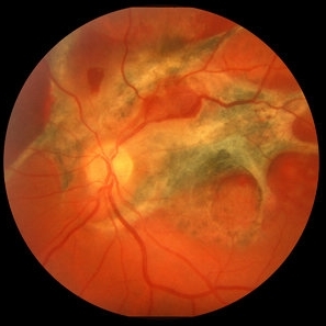
 Ocular Toxocariasis slide 1
Oct 22 2012 by Ronald C. Gentile, MD
40-year-old man from South America was referred for a peripheral retinal scar in his left eye. He had a history of conjunctivitis as a child with exposure to multiple pets (cats and dogs). Fundus photo revealed a peripheral scarred sub-retinal granuloma located superior nasal with a retinal fold and traction extending to the optic nerve.
Photographer: The New York Eye & Ear Infirmary Department of Medical Imaging
Condition/keywords: toxocariasis
-
 Disciform Scar
Jul 13 2013 by Jason S. Calhoun
Chorioretinal scar inferior temporal in the right eye of a middle aged patient.
Photographer: Jason S. Calhoun, Department of Ophthalmology, Mayo Clinic Jacksonville, Florida
Condition/keywords: chorioretinal scar
-
 Traumatic Chorioretinal Scarring
Oct 15 2012 by Jeffrey G. Gross, MD, FASRS
Traumatic chorioretinal scarring, with less hemorrhage, 1 month later.
Condition/keywords: chorioretinal scar
-
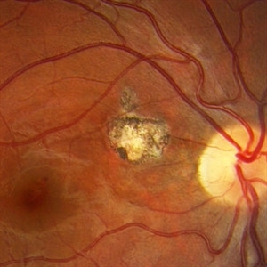
 Toxoplasma chorioretinitis 1
Jan 11 2013 by Alex P. Hunyor, MD
Toxoplasmosis 1 - chorioretinal scar from previous toxoplasma chorioretinitis. See image 2 - recurrent todo adjacent to this scar
Condition/keywords: inactive toxoplasmosis, ocular toxoplasmosis, toxoplasmosis, toxoplasmosis retinitis
-
 Chorioretinal Scar
Apr 1 2016 by Nichole Lewis
Chorioretinal scar.
Photographer: Nichole Lewis - Pennsylvania Retina Specialists, Camp Hill, PA
Condition/keywords: chorioretinal scar
-
---thumb.jpg/image-square;max$300,300.ImageHandler) Toxo Macular Scar
Toxo Macular Scar
Oct 15 2013 by Sjakon G Tahija, MD
Fundus photograph of a 22-year-old woman with a congenital choreoretinal scar from toxoplasma in the left eye. Vision in the right eye is 0.05. The right eye is NLP.
Condition/keywords: toxoplasmosis
-
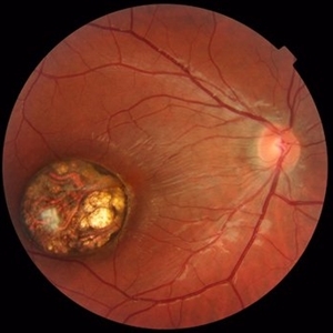
 Ocular Toxocariasis slide 3
Oct 22 2012 by Ronald C. Gentile, MD
The sub-retinal scarred granuloma was white in color and elevated. It had pigment speckling around it. Serum testing was positive for past exposure to Toxocara canis.
Photographer: The New York Eye & Ear Infirmary Department of Medical Imaging
Condition/keywords: toxocariasis
-
 Choroidal Hemangioma
Oct 20 2012 by Hyung-Woo Kwak, MD
Fundus, ICG, and OCT examination showed a typical chorioretinal scar lying concentric to the optic disc. Typical choroidal rupture was performed after intravitreal gas injection under the diagnosis of submacular hemorrhage caused by trauma, after the absorption of submacular hemorrhage
Condition/keywords: chorioretinal scar, choroidal rupture, submacular hemorrhage
-
---thumb.JPG/image-square;max$300,300.ImageHandler) Traumatic Optic Neuropathy
Traumatic Optic Neuropathy
Dec 9 2012 by Mallika Goyal, MD
Right eye of a 23-year-old gentleman 6 months following a road accident. Optic disc pallor with peripapillary chorioretinal scarring suggests traumatic optic neuropathy as the cause of optic atrophy.
Photographer: Mallika Goyal, MD, Apollo Health City, Hyderabad, India
Condition/keywords: traumatic optic neuropathy
-
 Congenital Toxoplasmosis
Oct 10 2015 by Hamid Ahmadieh, MD
Color fundus photograph of the right eye of a 15 -year-old boy with decreased vision due to a large chorioretinal scar involving the macula . The lesion is typical for a congenital ocular toxoplasmosis .
Photographer: Solmaz Shahmohammad, Negah Eye Center, Tehran, Iran
Condition/keywords: color fundus photograph, congenital toxoplasmosis
-
 Retinitis Sclopetaria, 6 months later
Jun 29 2018 by Gareth Lema, MD, PhD
Retinitis sclopetaria has resolved. There are multiple, large choroidal ruptures and subretinal scarring.
Photographer: Flaum Eye Institute, University of Rochester, Rochester, NY
Condition/keywords: blunt trauma, chorioretinitis sclopetaria
-
 Toxoplasmosis Slide 1
Oct 22 2012 by Ronald C. Gentile, MD
Focal, white area of chorioretinitis with overlying vitreous inflammation adjacent to an old chorioretinal scar in a patient complaining of photophobia, floaters and a decrease in vision of the right eye. The focal area of chorioretinitis is involving the inferior nasal macula and adjacent optic nerve with surrounding retinal and peri-papillary edema.
Photographer: The New York Eye & Ear Infirmary Department of Medical Imaging
Condition/keywords: posterior uveitis, toxoplasmosis
-
---thumb.jpg/image-square;max$300,300.ImageHandler) Old Presumed Ocular Histoplasmosis Syndrome
Old Presumed Ocular Histoplasmosis Syndrome
Feb 26 2013 by Henry J. Kaplan, MD
Old POHS, advanced subretinal scar formation due to CNV (end stage).
Condition/keywords: end stage, presumed ocular histoplasmosis syndrome (POHS), subretinal scar formation
-
 Toxoplasma Chorioretinal Scar
Mar 1 2014 by Homayoun Tabandeh, MD, FASRS
Toxoplasma chorioretinal scar.
Condition/keywords: toxoplasmosis chorioretinitis
-
 Toxoplasmosis Slide 1
Oct 22 2012 by Ronald C. Gentile, MD
35-year-old women presented with decreasing vision in the left eye with progressive central scotoma. Fundus examination revealed one focal area of chorioretinitis adjacent to one of multiple old pigmented retinal scars. The focal area of chorioretinitis involved the deep retinal layers and was associated with sub-retinal fluid and little overlying vitritis.
Photographer: The New York Eye & Ear Infirmary Department of Medical Imaging
Condition/keywords: punctate outer retinal toxoplasmosis, toxoplasmosis
-
---thumb.jpg/image-square;max$300,300.ImageHandler) Chorioretinal Scarring
Chorioretinal Scarring
Feb 15 2013 by From the Collections of Thomas M. Aaberg, MD and Thomas M. Aaberg Jr., MD
Color fundus photograph showing chorioretinal scarring consistent with prior retinal laser photocoagulation to areas of peripheral retinal nonperfusion.
Condition/keywords: laser scarring, peripheral retinal nonperfusion
-
---thumb.jpg/image-square;max$300,300.ImageHandler) Non-Specific Old Chorioretinetic Scars
Non-Specific Old Chorioretinetic Scars
Dec 5 2013 by Maurice F. Rabb
Non-specific old chorioretinetic scars.
Condition/keywords: chorioretinal scar
-
 Chorioretinal Scar
May 16 2017 by Olivia Rainey
Fundus photograph of an 17-year-old male with a macular scar affecting his right eye secondary to exudation from Coats disease.
Photographer: Olivia Rainey
Imaging device: Topcon 50dx
Condition/keywords: 20 degrees, chorioretinal scar, Coats' disease, color fundus photograph, color photo, fundus photograph
-
---thumb.jpg/image-square;max$300,300.ImageHandler) Non-Specific Old Chorioretinetic Scars
Non-Specific Old Chorioretinetic Scars
Dec 5 2013 by Maurice F. Rabb
Non-specific old chorioretinetic scars.
Condition/keywords: chorioretinal scar
-
---thumb.jpg/image-square;max$300,300.ImageHandler) Non-Specific Old Chorioretinetic Scars
Non-Specific Old Chorioretinetic Scars
Dec 5 2013 by Maurice F. Rabb
Non-specific old chorioretinetic scars.
Condition/keywords: chorioretinal scar
-
---thumb.jpg/image-square;max$300,300.ImageHandler) Non-Specific Old Chorioretinetic Scars
Non-Specific Old Chorioretinetic Scars
Dec 5 2013 by Maurice F. Rabb
Non-specific old chorioretinetic scars.
Condition/keywords: chorioretinal scar
-
 CR Scarring
Mar 17 2015 by Jason Griffith
Photograph of a 62 year old male with history of retinal detachment and resulting CR scarring.
Photographer: Jason Griffith, Tennessee Retina, Nashville, TN
Imaging device: Topcon TRC-50EX
Condition/keywords: chorioretinal scar
-
 Central Retinal Vein Occlusion
Jul 13 2018 by Olivia Rainey
Ultra-wide field, pseudocolor montage of a patient presenting with a central retinal vein occlusion, as well as, an inferior chorioretinal scar in their right eye.
Photographer: Olivia Rainey
Imaging device: Optos
Condition/keywords: central retinal vein occlusion (CRVO), chorioretinal scar, montage, Optos, pseudocolor, ultra-wide field imaging
-
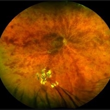
 multifocal choroiditis
Feb 14 2013 by From the Collections of Thomas M. Aaberg, MD and Thomas M. Aaberg Jr., MD
color fundus photos showing healed chorioretinal scars, pigment deposition, and subretinal fibrosis consistent with regressed multifocal choroiditis
Condition/keywords: multifocal choroiditis, posterior segment inflammation, subretinal fibrosis, white dot syndrome
-
 Disseminated Chorioretinitis With Unknown Etiology
Apr 5 2018 by Kim Barrett
Ultra-wide field fluorescein angiogram of a 31-year-old female with intermittent pain in her left eye. Her condition has been managed in Liberia until recently when she moved to the United States. She suffers from multiple modalities including central retinal artery occlusion, posterior synechiae of the iris, interstitial keratitis, disseminated chorioretinitis, as well as HIV. An infectious cause is high on the differential in light of her HIV status. DDx: hypertensive crisis, an embolism (? IV drug use), coagulopathy, trauma, infectious. Blood work was normal. Her current vision is 20/30 right eye and 20/400 left eye.
Photographer: Kim Barrett, COA
Imaging device: Optos
Condition/keywords: central retinal artery occlusion (CRAO), chorioretinal scar, ciliary artery sparring, disseminated chorioretinitis, HIV, left eye, optic atrophy, staining
 Loading…