Search results (24 results)
-
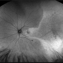
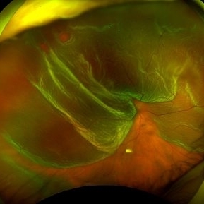
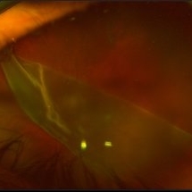
 Macula-Off Retinal Detachment
Jan 3 2017 by Jason Griffith
56-year-old female with "macula-off" retinal detachment.
Photographer: Jason Griffith
Imaging device: Topcon TRC-50EX
Condition/keywords: retinal detachment of the macula
-
---thumb.jpg/image-square;max$300,300.ImageHandler) Retinal Detachment Of The Macula
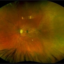
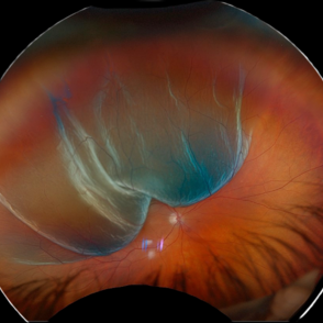
Retinal Detachment Of The Macula
Oct 24 2013 by Maurice F. Rabb
65 year old Caucasion male with a giant tear of the macula.
Condition/keywords: retinal detachment of the macula
-
---thumb.jpg/image-square;max$300,300.ImageHandler) Retinal Detachment Of The Macula
Retinal Detachment Of The Macula
Oct 24 2013 by Maurice F. Rabb
65 year old Caucasion male with a giant tear of the macula.
Condition/keywords: retinal detachment of the macula
-
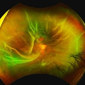
 Macula Off Retinal Detachment with CNV
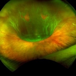
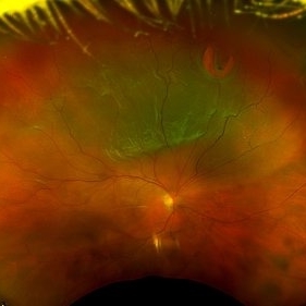
Nov 11 2019 by Olivia Rainey
Ultra-wide field pseudocolor photograph of a 42-year-old female with a long-standing, macula-off retinal detachment affecting her left eye. Patient was unaware of vision loss until testing her visual acuity and she denied seeing flashing lights. Patient decided to proceed with scleral buckling. The CNV is potentially secondary the retinal detachment, but may be myopic related or idiopathic. The CNV appears fibrotic and inactive. The patient was warned that this will absolutely limit how much vision she recovers once the retina is reattached.
Photographer: Olivia Rainey
Imaging device: Optos California
Condition/keywords: choroidal neovascularization (CNV), chronic retinal detachment, fundus autofluorescence (FAF), left eye, montage, Optos, retinal detachment of the macula, ultra-wide field imaging
-
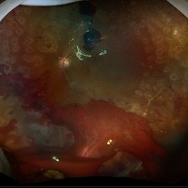
 Retinal Detachment
Aug 24 2015 by René Hernán Parada Vásquez
Fundus photograph of 52-year-old male with superior temporal retinal detachment.
Photographer: Parada René, ESO, Guatemala.
Imaging device: Canon CR-2
Condition/keywords: retinal detachment of the macula
-
 Macula Off Retinal Detachment with CNV
Nov 11 2019 by Olivia Rainey
Ultra-wide field pseudocolor photograph of a 42-year-old female with a long-standing, macula-off retinal detachment affecting her left eye. Patient was unaware of vision loss until testing her visual acuity and she denied seeing flashing lights. Patient decided to proceed with scleral buckling. The CNV is potentially secondary the retinal detachment, but may be myopic related or idiopathic. The CNV appears fibrotic and inactive. The patient was warned that this will absolutely limit how much vision she recovers once the retina is reattached.
Photographer: Olivia Rainey
Imaging device: Optos California
Condition/keywords: choroidal neovascularization (CNV), left eye, montage, Optos, pseudocolor, retinal detachment of the macula, ultra-wide field imaging
-
 RD Montage
Jul 3 2021 by Somnath Chakraborty, MD
Fundus photo montage of the left eye of a 56-year-old male showing subtotal retinal detachment with macular involvement and a large circumlinear tear extending from 1 o' clock to 3 o' clock hours.
Photographer: Pulak Roy
Condition/keywords: acute retinal detachment, retinal detachment of the macula, retinal tear, retinal tear with detachment
-
 Peripheral Retinoschisis
Feb 12 2020 by DIEGO TOLENTINO
Peripheral retinoschisis plus macula off retinal detachment.
Photographer: Diego Tolentino, CEOP
Condition/keywords: retinal detachment of the macula, senile retinoschisis
-
 Retinal Detachment with Tear
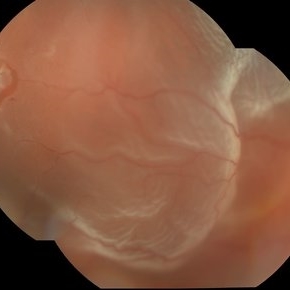
Jan 11 2022 by Manish Nagpal, MD, FRCS (UK), FASRS
Intraoperative photo of a retinal detachment with a posterior tear with everted edges.
Photographer: Manish Nagpal, Retinal Foundation, Ahmedabad, India
Imaging device: Sony PMW -10 MD surgical camera
Condition/keywords: retinal detachment of the macula, retinal detachment with retinal defect, retinal detachment with tear
-
 Retinal Detachment
Jan 12 2022 by Manish Nagpal, MD, FRCS (UK), FASRS
Intraoperative photo of a rhegmatogenous retinal detachment.
Photographer: Manish Nagpal, Retina Foundation, Ahmedabad, India
Imaging device: Sony PMW -10 MD surgical camera
Condition/keywords: retinal detachment of the macula
-
 Retinal Detachment
Oct 26 2023 by Virginia Gebhart
74 year old male with mac-off retinal detachment with single break
Photographer: Virginia Gebhart
Imaging device: Optos
Condition/keywords: detachment, Retinal Detachment, retinal detachment of the macula
-
 New Retinal Detachment 6w s/p RD repair
Nov 16 2023 by Virginia Gebhart
13 year old male presented with new blind spot 6 weeks s/p RD repair with cryo/scleral buckle/prophylaxis laser with gas bubble. New RD involving the macula, posterior to scleral buckle, secondary to PVD. Small gas bubble remaining. Pt was brought back to OR for repeat PPV and silicone oil repair
Photographer: Virginia Gebhart
Imaging device: Optos
Condition/keywords: gas bubble, Retinal Detachment, retinal detachment of the macula, scleral buckle
-
 Retinal Detachment with Multiple Retinal Tears
Jan 13 2021 by Kristen Wagner
Optos image of a male with a macular off retinal detachment and multiple retinal tears. This patient had detachment in both eyes.
Photographer: Kristen Wagner, COT Tennessee Retina Nashville TN
Condition/keywords: retinal detachment of the macula, retinal tear, retinal tear with detachment
-
 Mac off Retinal Detachment with Horseshoe Tear
Dec 5 2023 by Virginia Gebhart
68 year old male presented with HM vision in OD. Near total detachment with multiple breaks. Scheduled PPV with GFE. Visual prognosis guarded
Photographer: Virginia Gebhart
Imaging device: Topcon
Condition/keywords: Retinal Detachment, retinal detachment of the macula, Retinal Detachment with multiple breaks
-
 Macula off Retinal Detachment
Jan 23 2024 by Annaka Gooding
Ultra-widefield fundus photograph of an 81-year-old male with a Macula Off Retinal Detachment affecting his right eye. Patient presented at office with complaints of flashes of light for about 2 weeks accompanied by a curtain veil covering inferior visual field. Patient had total vision loss 24 hours prior to visit. His vision was scHM. The physician recommended Retinal Detachment Repair with PPV.
Photographer: Annaka Gooding, CPO
Imaging device: Optos California
Condition/keywords: detachment, fundus photography, macula off retinal detachment, Optos, retinal detachment of the macula, right eye, ultra-wide field imaging
-
 Morning glory disc anomaly-associated maculopathy: fibroglial tissue with a Mac-Off serous retinal detachment.
Jun 26 2024 by Julián Villarreal, MD
19 year old with a Morning glory disc anomaly-associated maculopathy: fibroglial tissue with a Mac-Off serous retinal detachment.
Photographer: Julián Villarreal MD
Imaging device: Mirante
Condition/keywords: fibroglial tissue, Morning Glory Anomaly, retinal detachment of the macula
-
 New RD with Multiple Breaks
Aug 26 2024 by Virginia Gebhart
59 year old male with superior, bullous, mac off RD with multiple breaks. Pt scheduled for PPV, laser, GFE.
Photographer: Virginia Gebhart
Imaging device: Optos California
Condition/keywords: retinal detachment of the macula, retinal tear, retinal tear with detachment
-
 Macula-Involving Rhegmatogenous Retinal Detachment
Feb 17 2024 by Nikhil K Bommakanti, MD
A middle-aged man with a history of rhegmatogenous retinal detachment repair in the fellow eye several years prior presented with reduced vision, which he had noticed two days before.
Condition/keywords: retinal detachment of the macula, rhegmatogenous retinal detachment
-
 Retinal Detachment with Single Break
May 30 2024 by Virginia Gebhart
73 year old female with new mac-off retinal detachment with single horseshoe tear. Vision CF@3ft. Repaired in OR with gas bubble and laser. Guarded prognosis
Photographer: Virginia Gebhart
Imaging device: Optos California
Condition/keywords: retinal detachment of the macula
-
 Retinal Detachment (Mac-Off)
Feb 20 2025 by Virginia Gebhart
63 year old male with a mac-off retinal detachment from 4:00 to 1:30 with a single break at 10:00. Pt schedule for PPV/GFE. Guarded prognosis for visual recovery.
Photographer: Virginia Gebhart, Retina Consultants of Carolina
Imaging device: Optos California
Condition/keywords: horseshoe tear, retinal detachment, retinal detachment of the macula
-
 Diabetic Tractional Retinal Detachment 1 week s/p SO fill
Aug 14 2024 by Virginia Gebhart
21 year old male 1 week s/p PPV/laser/STR/SO. Eye is stable, PRHs inferior and superior, possible traction from PRH/membrane. Will observe and let clot liquify, will consider scleral buckle if no improvement
Photographer: Virginia Gebhart
Imaging device: Optos California
Condition/keywords: Diabetic Tractional Detachment, retinal detachment of the macula, silicone oil
-
 Retinal Detachment with Giant Retinal Tear
Mar 26 2024 by Xitlali Caterina
Ultra-widefield fundus photograph of a 43-year-old male with a Retinal Detachment with Giant Retinal Tear affecting his left eye. Patient presented to the office with count fingers vision at 2 feet. He stated that about 8-9 days ago, he developed a clear curtain/veil and his vision started to get blurry. He also noted that he had floaters and flashes for about 8-9 days as well. The patient had cataract surgery a month prior to his visit. He stated that since his surgery, his vision had been better, but he had an area where he was not able to see well. The physician recommended a complex retinal detachment repair.
Photographer: Xitlali Caterina
Imaging device: OPTOS California RGB
Condition/keywords: fundus photograph, giant retinal tear, left eye, Optos, OPTOS CALIFORNIA, retinal detachment of the macula, retinal detachment with tear, ultra-wide field imaging, ultra-widefield image
-
 Rhegmatogenous Retinal Detachment
Mar 24 2025 by DR APOORVA JADHAV, MBBS , DNB
This is a color fundus photograph showing rhegmatogenous retinal detachment with posterior pole retinal tear with macula off.
Condition/keywords: retinal detachment of the macula
-
 Bullous Retinal Detachment
Nov 13 2025 by Virginia Gebhart
42 year old female referred for vision loss x 4-5 days. Bullous retinal detachment from 8:00 to 3:00 with retinal tear at 11:00. Macula is detached. Vision is LP, IOP of 3. Pt is scheduled for GFE and possible scleral buckle.
Photographer: Virginia Gebhart, Retina Consultants of Carolina
Imaging device: Optos California
Condition/keywords: bullous retinal detachment, retinal detachment, retinal detachment of the macula, retinal tear with detachment
 Loading…