Search results (126 results)
-
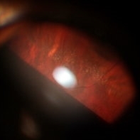
 Operculated Retinal Hole
Oct 12 2012 by Jeffrey G. Gross, MD, FASRS
Operculated retinal hole with RD.
Condition/keywords: operculated retinal hole, retinal degeneration
-
 Snail Track Peripheral Retinal Degeneration
Apr 29 2022 by Otakar Dušek, M.D. Ph.D.
Colour fundus photograph of 22-year-old woman with incidentally found snail track retinal degeneration in the superior temporal periphery of the retina of the right eye.
Photographer: Otakar Dušek, Charles University, Prague
Imaging device: Zeiss Clarus
Condition/keywords: peripheral retinal degeneration
-
 Operculated Retinal Hole in Retinal Detachment
Oct 12 2012 by Jeffrey G. Gross, MD, FASRS
Operculated retinal hole in retinal detachment.
Condition/keywords: operculated retinal hole, retinal degeneration
-
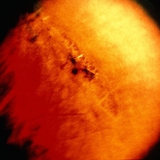
 Giant Retinal Tear
Oct 9 2012 by Audina M. Berrocal, MD FASRS
Teenager with high myopia and a GRT
Photographer: Ditte Hess CRA, BPEI
Imaging device: Fundus Camera
Condition/keywords: high myopia, retinal degeneration, retinal tear
-
 Retinal Detachment with Macula Partially Detached
Oct 12 2012 by Jeffrey G. Gross, MD, FASRS
RD with macula partially detached.
Condition/keywords: macula, retinal degeneration
-
 Pigmented Peripheral Retinal Degeneration
Jun 27 2013 by Jason S. Calhoun
42-year-old male came in for routine eye exam and to follow up on peripheral retinal degeneration in both eyes. VA is 20/20, right eye and 20/25, left eye. Patient is asymptomatic with no visual complaints.
Photographer: Jason S. Calhoun, Mayo Clinic Jacksonville, Florida
Imaging device: TOPCON TRC 50-EX
Condition/keywords: peripheral retinal degeneration
-
---thumb.JPG/image-square;max$300,300.ImageHandler) Peripheral Retinal Degeneration
Peripheral Retinal Degeneration
Jul 8 2013 by Jason S. Calhoun
Patient comes in with double vision. VA was 20/20 in both eyes. Fundus exam shows retinal degenerative changes in both eyes. Offer to correct double vision with temporary Fresnel prism.
Photographer: Jason S. Calhoun, Department of Ophthalmology, Mayo Clinic Jacksonville, Florida
Condition/keywords: peripheral retinal degeneration
-
 Lattice Degeneration
May 2 2013 by Henry J. Kaplan, MD
Pigmented lattice degeneration with lattice "wicker" caused by sclerotic blood vessels.
Condition/keywords: lattice degeneration, peripheral retinal degeneration
-
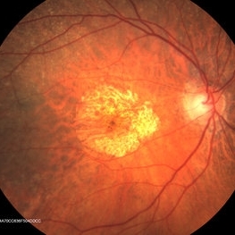 ARMD With Geographic Atrophy, Peripheral Degeneration
Dec 6 2013 by James B. Soque, CRA, OCT-C, COA, FOPS
92-year-old white female with exudative macular degeneration, geographic atrophy, and peripheral retinal degeneration.
Photographer: James Soque, CRA COA, Island Retina, Shirley, New York
Imaging device: Topcon TRC 50DX with OIS 10.6.45
Condition/keywords: geographic atrophy, macular degeneration, retinal degeneration
-
 Rhegmatogenous Retinal Detachment
Oct 5 2012 by Ronald C. Gentile, MD
Rhegmatogenous retinal detachment (right eye, temporal retina) involving the macula with characteristic outer hydration lines.
Photographer: The New York Eye & Ear Infirmary Department of Medical Imaging
Condition/keywords: retinal degeneration
-
 Rhegmatogenous Retinal Detachment
Oct 5 2012 by Ronald C. Gentile, MD
Rhegmatogenous retinal detachment involving the macula with characteristic outer hydration lines and associated peripheral retinal tear (top left of image). The choroidal vasculature can be seen through the break in the retina.
Photographer: The New York Eye & Ear Infirmary Department of Medical Imaging
Condition/keywords: retinal degeneration
-
 Macula-Sparing GRT RRD
Jul 6 2017 by Andrew A. Moshfeghi, MD, MBA, FASRS
Wide-field fundus photograph of a 43-year-old myopic man with a history of lattice retinal degeneration status posterior barrier laser performed elsewhere who presented with a giant-retinal tear associated retinal detachment of the right eye.
Photographer: Jay Jiang, University of Southern California Roski Eye Institute
Imaging device: Optos California
Condition/keywords: acute retinal detachment, giant retinal tear, lattice degeneration
-
 Retinal Detachment
Nov 9 2012 by Norman Byer
This is a retinal detachment in a 55-year-old man. The vertical convex line on the right side probably represents the posterior border of the vitreous base. Note the small tractional tear with the base of its flap attached at this line. This demonstrates how the vitreous base presents an effective barrier to further extension of the retinal tear. Note also how the flap breaks the continuity of the yellow line.
Condition/keywords: retinal degeneration, retinal flap, tractional retinal tear, vitreous base
-
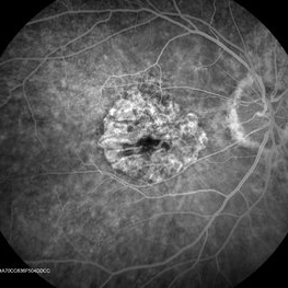 ARMD With Geographic Atrophy , Peripheral Degeneration, FA
Dec 6 2013 by James B. Soque, CRA, OCT-C, COA, FOPS
FA right eye early phase, of a 92-year-old white female with exudative macular degeneration, geographic atrophy, and peripheral retinal degeneration.
Photographer: James Soque CRA COA, Island Retina, Shirley, New York
Imaging device: Topcon TRC 50DX with OIS 10.6.45
Condition/keywords: geographic atrophy
-
 Stickler Syndrome
Dec 8 2016 by Aleksandra V. Rachitskaya, MD, FASRS
Optos wide-field fundus image of a patient with Stickler Syndrome and COL2A1 gene mutation. Patient has perviously undergone prophylactic laser. Lattice, vitreous veils, and laser scars are seen.
Photographer: Anne Pinter, Cole Eye Institute, Cleveland Clinic
Condition/keywords: Stickler Syndrome, vitreoretinal degeneration
-
 ARMD With Geographic Atrophy, Peripheral Degeneration
Dec 6 2013 by James B. Soque, CRA, OCT-C, COA, FOPS
92-year-old white female with exudative macular degeneration, geographic atrophy, and peripheral retinal degeneration.
Photographer: James Soque, CRA COA, Island Retina, Shirley, New York
Imaging device: Topcon TRC 50DX with OIS 10.6.45
Condition/keywords: fundus photograph, geographic atrophy
-
 Peripheral Retinal Degeneration
Jul 8 2013 by Jason S. Calhoun
Patient in with double vision. VA was 20/20 in both eyes. Fundus exam shows retinal degenerative changes in both eyes. Offer to correct double vision with temporary Fresnel prism.
Photographer: Jason S. Calhoun, Department of Ophthalmology, Mayo Clinic Jacksonville, Florida
Condition/keywords: peripheral retinal degeneration
-
 Snowflake Vitreoretinal Degeneration
Nov 29 2018 by Hashim Ali Khan, OD, FAAO
Peripheral snowflake in a 16-year-old female. The fellow eye had chronic total retinal detachment.
Imaging device: Goldman triple mirror lens
Condition/keywords: peripheral fundus lesion, snowflake hereditary degeneration
-
 Unusual Tapetoretinal Degeneration
Mar 14 2014 by David Callanan, MD
13-year-old black male with unusual tapetoretinal degeneration.
Condition/keywords: tapeoretinal degeneration
-
 Pigmented Paravenous Chorioretinal Atrophy
Feb 1 2018 by John S. King, MD
15-year-old healthy, asymptomatic AAM; found on routine eye exam; no FHx of RP known; OCT shows some extrafoveal, outer retinal degeneration.
Photographer: Karin Aletter
Condition/keywords: pigmented paravenous chorioretinal atrophy (PPCRA)
-
---thumb.jpg/image-square;max$300,300.ImageHandler) RD Repair Cartoon
RD Repair Cartoon
Feb 13 2013 by From the Collections of Thomas M. Aaberg, MD and Thomas M. Aaberg Jr., MD
Extrusion needle, endoilluminator.
Condition/keywords: cartoon, enclosed ora bay, extrusion needle, retinal degeneration
-
 Pigmented Paravenous Chorioretinal Atrophy
Feb 1 2018 by John S. King, MD
15 yo healthy, asymptomatic AAM; found on routine eye exam; no FHx of RP known; OCT shows some extrafoveal, outer retinal degeneration.
Photographer: Karin Aletter
Condition/keywords: pigmented paravenous chorioretinal atrophy (PPCRA)
-
 Pigmented Paravenous Chorioretinal Atrophy
Feb 1 2018 by John S. King, MD
15-year-old healthy, asymptomatic AAM; found on routine eye exam; no FHx of RP known; OCT shows some extrafoveal, outer retinal degeneration.
Photographer: Karin Aletter
Condition/keywords: pigmented paravenous chorioretinal atrophy (PPCRA)
-
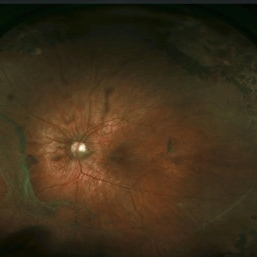
 Macular Hole Retinal Detachment Over a Posterior Staphyloma
Dec 31 2016 by Linda A Cernichiaro- Espinosa, MD
Macular hole retinal detachment over a posterior staphyloma of pathologic myopia.
Photographer: Linda A Cernichiaro
Imaging device: Optos
Condition/keywords: degenerative myopia, high myopia, macular hole, myopic eye, posterior staphyloma, vitreoretinal degeneration
-
---thumb.jpg/image-square;max$300,300.ImageHandler) Retinal Degeneration
Retinal Degeneration
Oct 7 2013 by Maurice F. Rabb
Nine year old Afro American girl who was referred for the evaluation of a retinal degeneration. Visual acuity was correctable to 20/60 in each eye. Intraocular pressures where 13 mmHg OD and 12 mmHg OS. There was a trace to 1 + cells in the right vitreous and +1 to +2 cells in the left. Fundus exam showed evidence for diffuse RPE hypopigmentation. The changes were consistent with a diffuse pigmentary degeneration of the retina in each eye. The patient's retinal degeneration is associated with a systemic syndrome.
Condition/keywords: retinal degeneration
 Loading…