Search results (433 results)
-
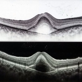
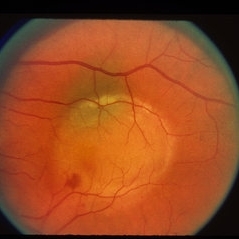
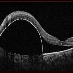
 PED due to CSCR
Sep 2 2012 by Hamid Ahmadieh, MD
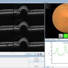
OCT image of a 37-year-old man with a serous PED secondary to CSCR.
Photographer: Hamid Ahmadieh, Ophthalmic Research Center, Labbafinejad Medical Center
Imaging device: Heidelberg Spectralis
Condition/keywords: central serous chorioretinopathy (CSCR), optical coherence tomography (OCT), pigment epithelial detachment (PED)
-
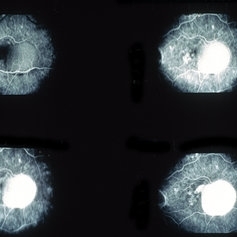
 Pigment Epithelial Detachment late FA with small occult CNV
Jul 6 2012 by Tarek S. Hassan, MD, FASRS
72-year-old man with VA loss and metamorphopsia of 2 months duration. PED found, testing done to rule out CNV. Very suspicious for CNV in superonasal fovea/parafovea.
Condition/keywords: choroidal neovascularization (CNV), pigment epithelial detachment (PED)
-
 Spontaneous Flattening of Drusenoid PED
Jul 1 2014 by John S. King, MD
Consult to r/o ExAMD; observed; scans about a year apart.
Photographer: Wayne A Ladlee Jr
Imaging device: Cirrus
Condition/keywords: drusenoid PED, macular drusenoid lesion, pigment epithelial detachment (PED)
-
 Fibrovascular PED
Feb 21 2014 by Roy Schwartz, MD
72-year-old female with fibrovascular PED. Upper picture - PED with sub RPE hyper-reflective substance, in a multi-layered pattern, corresponding to fibrovascular PED. CME. Lower picture - PED flattened, a denser sub RPE hyperreflective substance is seen. CME resolved.
Condition/keywords: fibrovascular pigment epithelial detachment (PED), neovascular age-related macular degeneration (AMD), optical coherence tomography (OCT), ranibizumab
-
 Subfoveal Choroidal Neovascularization, In Stereo
Sep 28 2012 by Michael P. Kelly, FOPS
Subfoveal Choroidal Neovascularization.
Photographer: Michael P. Kelly, FOPS Director, Duke Eye Labs, Duke University Hospital, Duke Eye Center, Durham, NC
Imaging device: Zeiss FF4
Condition/keywords: pigment epithelial detachment (PED), stereo pair, subfoveal choroidal neovascularization
-
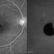
 PED due to CSCR 4
Sep 2 2012 by Hamid Ahmadieh, MD
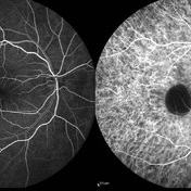
Early phase FA & ICG images of a 37-year-old man with a serous PED secondary to CSCR
Photographer: Hamid Ahmadieh, Ophthalmic Research Center, Labbafinejad Medical Center
Imaging device: Heidelberg Spectralis
Condition/keywords: central serous chorioretinopathy (CSCR), indocyanine green (ICG) angiography, pigment epithelial detachment (PED)
-
 Fibrovascular Retinal Pigment Epithelial Detachment - Color Fundus
Jul 16 2014 by James B. Soque, CRA, OCT-C, COA, FOPS
69-year-old white female with Hx of 10 anti-VEFG treatment injections of right eye, VA 20/200, now stable, off drug for 10 months.
Photographer: James B Soque, CRA COA
Imaging device: Topcon TRC 50 DX with MERGE software, 5 MP dig camera
Condition/keywords: color fundus photograph, fibrovascular pigment epithelial detachment (PED), pigment epithelial atrophy, retina
-
 PED due to CSCR 2
Sep 2 2012 by Hamid Ahmadieh, MD
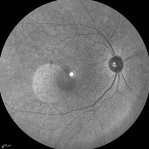
Autofluorescence imaging of a 37-year-old man with a serous PED secondary to CSCR.
Photographer: Hamid Ahmadieh, Ophthalmic Research Center, Labbafinejad Medical Center
Imaging device: Heidelberg Spectralis
Condition/keywords: autofluorescence imaging, central serous chorioretinopathy (CSCR), pigment epithelial detachment (PED)
-
 Pigment Epithelial Detachment
Jul 6 2012 by Tarek S. Hassan, MD, FASRS
72-year-old man with VA loss and metamorphopsia of 2 months duration. PED found, testing done to rule out CNV.
Condition/keywords: metamorphopsia, pigment epithelial detachment (PED)
-
 Vogt-Koyanagi-Harada with Multiple PEDs
Oct 10 2012 by Jeffrey G. Gross, MD, FASRS
VKH with multiple PEDs, FA mid phase.
Condition/keywords: FA mid phase, pigment epithelial detachment (PED)
-
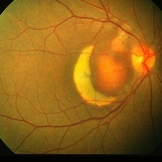
 Central Serous Chorioretinopathy (CSC)
Oct 16 2012 by S. Natarajan, MD, FASRS, FRCS (GLASGOW) , FICO, D.Sc, FELA
Middle-aged male came with small PED 4 months back; now this has progressed to a larger PED with SRF underneath the fovea.
Photographer: Prof. Dr. S. Natarajan
Condition/keywords: central serous chorioretinopathy (CSCR), central serous retinopathy (CSR), pigment epithelial detachment (PED), subretinal fibrosis
-
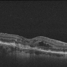
 Hemorrhagic Pigment Epithelial Detachment
Dec 14 2016 by Hashim Ali Khan, OD, FAAO
OCT of a 20-year-old female after trauma with tennis-ball, showing a hemorrhagic PED. RPE is elevated. The second Hyper-reflective band corresponding to Bruchs membrane (BM complex) is visible.
Condition/keywords: pigment epithelial detachment (PED), subretinal hemorrhage
-
 PED due to CSCR 5
Sep 2 2012 by Hamid Ahmadieh, MD
Late-phase FA and ICG images of a 37-year-old man with a serous PED secondary to CSCR.
Photographer: Hamid Ahmadieh, Ophthalmic Research Center, Labbafinejad Medical Center
Imaging device: Heidelberg Spectralis
Condition/keywords: central serous chorioretinopathy (CSCR), indocyanine green (ICG) angiography, pigment epithelial detachment (PED)
-
 PED due to CSCR 3
Sep 2 2012 by Hamid Ahmadieh, MD
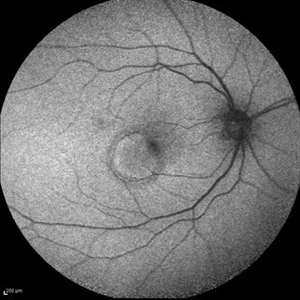
Infrared imaging of a 37-year-old man with a serous PED secondary to CSCR.
Photographer: Hamid Ahmadieh, Ophthalmic Research Center, Labbafinejad Medical Center
Imaging device: Heidelberg Spectralis
Condition/keywords: central serous chorioretinopathy (CSCR), pigment epithelial detachment (PED)
-
 Vogt-Koyanagi-Harada with Multiple PEDS
Oct 10 2012 by Jeffrey G. Gross, MD, FASRS
VKH, with multiple PEDs.
Condition/keywords: pigment epithelial detachment (PED)
-
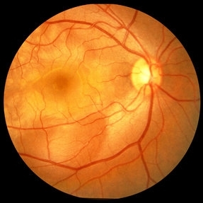
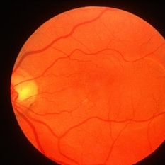
 Neurosensory Detachment with associated Pigment Epithelial Detachment
Oct 10 2012 by K. Bailey Freund, MD
Fundus photograph of a 39-year-old man with central serous chorioretinopathy noted to have a neurosensory detachment with associated pigment epithelial detachment.
Condition/keywords: central serous chorioretinopathy (CSCR), pigment epithelial detachment (PED)
-
 Fibrovascular PED
Jun 4 2014 by Henry J. Kaplan, MD
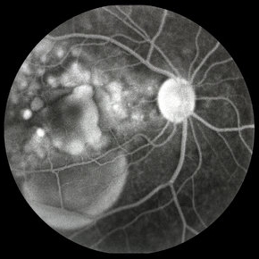
Fundus photograph of a large fibrovascular pigment epithelial detachment with inferonasal hemorrhage. #1
Condition/keywords: pigment epithelial detachment (PED), vascularized pigment epithelial detachment (PED)
-
 Fibrovascular PED
May 2 2013 by Henry J. Kaplan, MD
Fluorescein angiogram of the fibrovascular PED in the same patient; early homogeneous hyperfluorescence in the PED area which is increased in fluorescence to the late phase with a granular hyperfluorescence adjacent to PED in the foveal side which starts in the mid-phase of the F/A and is increased later; #2.
Condition/keywords: exudative age-related macular degeneration, fibrovascular pigment epithelial detachment (PED)
-
 Pigment Epithelial Detachment With Rip
Sep 4 2013 by Christopher T Cessna, DO
Fundus photograph of 76-year-old woman with exudative macular degeneration, PED with rip.
Photographer: Denise Miller, RD
Condition/keywords: macular degeneration, pigment epithelial detachment (PED)
-
 RPE rip macular OCT
Dec 23 2012 by Alex P. Hunyor, MD
80-year-old female with subfoveal occult CNV and large extrafoveal PED which underwent spontaneous RPE rip. OCT shows subfoveal CNV and intraretinal cystic edema
Condition/keywords: pigment epithelial detachment (PED), retinal pigment epithelium (RPE) tear
-
 Fibrovascular PED
May 2 2013 by Henry J. Kaplan, MD
Fundus photograph and fluorescein angiography of a fibrovascular PED with a typical notch on F/A.
Condition/keywords: exudative age-related macular degeneration, fibrovascular pigment epithelial detachment (PED), vascularized pigment epithelial detachment (PED)
-
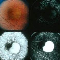
 Pigment Epithelial Detachment
Nov 20 2016 by JEFFERSON R SOUSA, Tecg.º (Biomedical Systems Technology)
Female patient, 43-years-old, Caucasian. Attended the clinic with complaint of low vision. In the fundus evaluation, DEP was observed in the upper region with macular involvement.
Photographer: JEFFERSON R SOUSA - Study Center and Ophthalmological Research Dr. Andre M V Gomes, Institute Dr. Suel Abujamra São Paulo-Brazil
Imaging device: OCT Cirrus - Zeiss / Cut in line from 11 to 5hr.
Condition/keywords: large pigment epithelial detachment, pigment epithelial detachment (PED)
-
 Fibrovascular Retinal Pigment Epithelial Detachment - Fluorescein Angiography
Jul 16 2014 by James B. Soque, CRA, OCT-C, COA, FOPS
69-year-old white female with Hx of 10 anti-VEFG treatment injections of right eye, VA 20/200, now stable, off drug for 10 months.
Photographer: James B Soque, CRA COA
Imaging device: Topcon TRC 50 DX with MERGE software, 5 MP dig camera
Condition/keywords: anti-VEGF, detachment, fibrovascular pigment epithelial detachment (PED), retinal pigment epithelium
-
 Fibrovascular PED
May 2 2013 by Henry J. Kaplan, MD
Elevated PED is visible in the temporal part of the macula; #1.
Condition/keywords: exudative age-related macular degeneration, fibrovascular pigment epithelial detachment (PED)
-
 Exudative AMD
May 2 2013 by Henry J. Kaplan, MD
Exudative AMD as hemorrhagic PED with adjacent old subretinal hemorrhage which has turned yellow in color.
Condition/keywords: exudative age-related macular degeneration, hemorrhagic PED, pigment epithelial detachment (PED)
 Loading…