Search results (37 results)
-
 Lacquer Cracks in Pathologic Myopia
Apr 19 2013 by Theodore Leng, MD, MS, FASRS
Color fundus photograph of a 48-year-old woman with lacquer cracks in the setting of pathologic myopia.
Condition/keywords: lacquer cracks, pathologic myopia
-
 Fuch's Spot
Apr 2 2019 by Gary R. Cook, MD, FACS
20-year-old patient with high myopia and a Fuch's spot OD.
Condition/keywords: Fuchs, high myopia, pathologic myopia
-
 OCT Myopic Staphyloma With Schisis and ERM
Apr 24 2014 by Scott E. Pautler, MD
OCT of high myope with asymptomatic macular schisis.
Imaging device: Heidelberg Spectralis
Condition/keywords: foveal schisis, maculopathy, maculoschisis, optical coherence tomography (OCT), pathologic myopia, staphyloma
-
 Lacquer Cracks in Pathologic Myopia
Apr 19 2013 by Theodore Leng, MD, MS, FASRS
Red free fundus photograph of a 48-year-old woman with lacquer cracks in the setting of pathologic myopia.
Condition/keywords: lacquer cracks, pathologic myopia
-
 Optos Picture With Speculum: Dislocated Natural Lens
Oct 9 2018 by John S. King, MD
55-year-old white female with history of pathologic myopia+, lattice (laser), SB OU (1990s), and dislocated natural lenses OU that had been watched for years. In the fellow eye she developed phacolytic glaucoma and a PPV, PPL was performed. Plan for both eyes are monitoring. I wanted to get a good picture of her lens today with the optos machine, as the other pics had artifact from the lower lid. It worked out well to use a speculum in the left eye. Vision cc is 20/400 J1+ OD and 20/40 J2 OS; aphakic OU; vitreous clear OD; dislocated lens OS (see pic); retinas attached.
Photographer: Maisee Yang
Imaging device: Optos California
Condition/keywords: dislocated crystalline lens, pathologic myopia, scleral buckle, staphyloma
-
 Laser Barrage for Temporal Localized Rhematogenous Retinal Detachment
Feb 15 2018 by Kushal S Delhiwala, MBBS, MS, FMRF,FICO, FAICO
39-year-old female presenting with sudden onset flashes and floaters in left eye having undergone refractive surgery 20 years before for pathologic myopia.Color fundus photograph montage of left eye showing macula sparing inferotemporal localized Rhematogenous retinal detachment with horse shoe tear and temporal lattice degeneration treated with laser barrage.
Photographer: Dr Kushal Delhiwala, Netralaya superspeciality eye hospital ,Ahmedabad
Imaging device: Zeiss Visucam 500
Condition/keywords: barrier laser, macula sparring
-
 High Myopia
Apr 2 2019 by Gary R. Cook, MD, FACS
17-year-old Vietnamese male with -23D myopia; V.A.= 20/40
Imaging device: Topcon VT-50
Condition/keywords: high myopia, lacquer cracks, pathologic myopia
-
 Myopic Traction Maculopathy
May 31 2014 by Rameez N Hussain, MD
Spectral domain optical coherence tomography of macular detachment in posterior staphyloma - myopic traction maculopathy (MTM).
Photographer: Rameez N Hussain MD, Vitreo Retinal Services, Giridhar Eye Institute, Cochin, India
Imaging device: Heidelberg Spectralis
Condition/keywords: high myopia, macular detachment, myopic traction maculopathy, pathologic myopia, posterior staphyloma
-
 Myopic Traction Maculopathy
May 31 2014 by Rameez N Hussain, MD
Color photograph of macular detachment in a posterior staphyloma - myopic traction maculopathy (MTM).
Photographer: Rameez N Hussain MD, Vitreo Retinal Services, Giridhar Eye Institute, Cochin, India
Imaging device: Zeiss
Condition/keywords: high myopia, macular detachment, myopic traction maculopathy, pathologic myopia, posterior staphyloma
-
 Myopic Degeneration
Jul 3 2018 by Armando L. Oliver, MD
Myopic Degeneration
Photographer: Moises Castro
Imaging device: Optos California
Condition/keywords: pathologic myopia, posterior staphyloma
-
 Macular Hole Retinal Detachment Over a Posterior Staphyloma
Dec 31 2016 by Linda A Cernichiaro- Espinosa, MD
Macular hole retinal detachment over a posterior staphyloma of pathologic myopia.
Photographer: Linda A Cernichiaro
Imaging device: Optos
Condition/keywords: degenerative myopia, high myopia, macular hole, myopic eye, posterior staphyloma, vitreoretinal degeneration
-
 High Myopia
Apr 2 2019 by Gary R. Cook, MD, FACS
51-year-old white female with -7.00D myopia with a myopic conus on temporal aspect of the optic nerve and focal choroiretinal atrophy in the macula OS; V.A. = 20/25-1
Imaging device: Topcon VT-50
Condition/keywords: high myopia, myopic degeneration, myopic fundus, pathologic myopia
-
 Myopic Degeneration
Jul 3 2018 by Armando L. Oliver, MD
Myopic Degeneration
Photographer: Moises Castro
Imaging device: Optos California
Condition/keywords: pathologic myopia, posterior staphyloma
-
 Myopic Degeneration
Jul 3 2018 by Armando L. Oliver, MD
Late Views IVFA
Photographer: Moises Castro
Imaging device: Optos California
Condition/keywords: pathologic myopia, posterior staphyloma
-
 Myopic Degeneration
Jul 3 2018 by Armando L. Oliver, MD
FAF
Photographer: Moises Castro
Imaging device: Optos California
Condition/keywords: pathologic myopia, posterior staphyloma
-
 Myopic Degeneration
Jul 3 2018 by Armando L. Oliver, MD
FAF
Photographer: Moises Castro
Imaging device: Optos California
Condition/keywords: pathologic myopia, posterior staphyloma
-
 High Myopia
Apr 2 2019 by Gary R. Cook, MD, FACS
51-year-old white female with -6.25D myopia OD with a myopic conus on the inferotemporal aspect of the optic disc and focal myopic chorioretinal atrophy in the macula OD; V.A. = 20/25
Imaging device: Topcon VT-50
Condition/keywords: high myopia, myopic degeneration, myopic fundus, pathologic myopia
-
 Myopic Degeneration
Jul 3 2018 by Armando L. Oliver, MD
Late views IVFA.
Photographer: Moises Castro
Imaging device: Optos California
Condition/keywords: pathologic myopia, posterior staphyloma
-
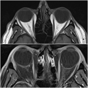 Staphyloma in Pathologic Myopia
Feb 7 2020 by Jonathan C. Tsui, MD
A 51-year-old presents with a six-month history of OS vision loss found to be CF at 3'. Fundus exam demonstrated pathologic myopia OS>OD with tilted discs. MRI Orbit Axial T1 images demonstrate significant compatible findings of staphylomas OS>OD.
Condition/keywords: pathologic myopia, staphyloma
-
 Bilateral CNV in High Myopia
Apr 2 2019 by Gary R. Cook, MD, FACS
Right eye of a 60-year-old white female with -9D myopia, myopic maculopathy, and visible (Type 1) CNV; V.A. = 20/40.
Imaging device: Topcon VT-50
Condition/keywords: choroidal neovascular membrane (CNVM), choroidal neovascularization (CNV), high myopia, myopic degeneration, myopic fundus, pathologic myopia
-
 Bilateral CNV in High Myopia
Apr 2 2019 by Gary R. Cook, MD, FACS
Left eye of a 60-year-old white female with -9D myopia and bilateral visible (Type 1) CNV; V.A. = 20/30.
Imaging device: Topcon VT-50
Condition/keywords: choroidal neovascular membrane (CNVM), choroidal neovascularization (CNV), high myopia, myopic degeneration, myopic fundus, pathologic myopia
-
 High Myopia with CNVM
Apr 2 2019 by Gary R. Cook, MD, FACS
61-year-old patient with -8.25D myopia and a type 1 CNVM OD; V.A. = 20/200
Imaging device: Topcon VT-50
Condition/keywords: choroidal neovascular membrane (CNVM), high myopia, pathologic myopia, subretinal neovascular membrane
-
 Visible Myopic CNVM
Apr 1 2019 by Gary R. Cook, MD, FACS
70-year-old white male with visible myopic CNVM OS; V.A.= 20/200.
Imaging device: Topcon VT-50
Condition/keywords: high myopia, myopic choroidal neovascularization (CNV), pathologic myopia
-
 CNVM due to Pathologic Myopia
Apr 2 2019 by Gary R. Cook, MD, FACS
55-year-old Asian male with -9.50D myopia with a visible (Type I) CNVM and thin hemorrhage in the macula: V.A.= 20/200
Imaging device: Topcon VT-50
Condition/keywords: choroidal neovascular membrane (CNVM), high myopia, pathologic myopia, retinal hemorrhage
-
 Degenerative Myopia
Apr 12 2023 by Ahmed Abbas Hashmi, OD
Right eye Fundus photograph of a 61-year-old female with pathological myopia.
Condition/keywords: chorioretinal atrophy, high myopia, pathologic myopia
 Loading…