Search results (52 results)
-
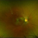
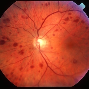 Ocular ischaemic syndrome colour 1
Jan 11 2013 by Alex P. Hunyor, MD
Ocular ischaemic syndrome, left eye - color image, posterior pole. Note: dilated but not tortuous veins, attenuated arteries, and multiple intraretinal haemorrhages.
Condition/keywords: ocular ischemic syndrome
-
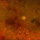
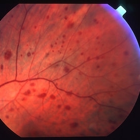 Ocular ischaemic syndrome colour 2
Jan 11 2013 by Alex P. Hunyor, MD
Ocular ischaemic syndrome, left eye - color image, superotemporal midperiphery. Note: dilated but not tortuous veins, attenuated arteries, and multiple intraretinal haemorrhages.
Condition/keywords: ocular ischemic syndrome
-
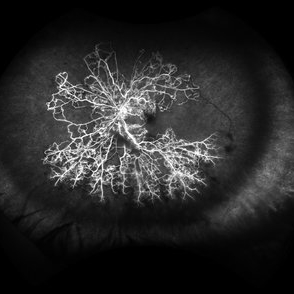
 Ocualr Ischemic Syndrome
Sep 22 2012 by Hamid Ahmadieh, MD
Wide field fluorescein angiography of a 66-year-old man with ocular ischemic syndrome due to severe stenosis of the right internal carotid artery.
Photographer: Hamid Ahmadieh, MD, Ophthalmic Research Center, Labbafinejad Medical Center, Shahid Beheshti University of Medical Sciences
Imaging device: HRA
Condition/keywords: carotid artery occlusion, ocular ischemic syndrome
-
 Ocular ischaemic syndrome FA 2
Jan 11 2013 by Alex P. Hunyor, MD
Ocular ischaemic syndrome, left eye - fluorescein angiogram, late phase.
Condition/keywords: ocular ischemic syndrome
-
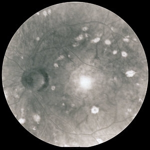
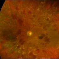
 Ocular Ischemic Syndrome
Jun 20 2018 by Andreas Ebneter, MD, PhD, FASRS
Ocular ischemic syndrome can present with a wide variety of ocular findings in both the anterior and posterior segments. The color fundus image of this 77-year-old male shows scattered blot hemorrhages in the deep retinal layers of the posterior pole that are only occasionally confluent. Commonly, these typical hemorrhages are predominantly found in the mid-periphery. Fluorescein angiography helps in confirming the diagnosis. Choroidal filling time is frequently somewhat delayed and patchy. Arteriovenous transit time is clearly prolonged. Staining of both veins and arteries in late images (top right) reflects diffuse endothelial cell damage with compromise of the blood-retina barrier. The peripheral retina is affected by extensive non-perfusion.
Photographer: Eva Steffen, Bern University Hospital, Switzerland
Imaging device: Optos 200Tx and Heidelberg Spectralis OCT
Condition/keywords: ocular ischemic syndrome
-
 Ocular Ischemic Syndrome
Aug 21 2020 by Gabriel Costa Andrade, PhD
Wide field fluorescein angiography of a 76-year-old male with ocular ischemic syndrome associated with severe stenosis of the left internal carotid artery.
Photographer: Gabriel Andrade
Imaging device: OPTOS - CALIFORNIA
Condition/keywords: ocular ischemic syndrome
-
 Scleritis / OIS
Oct 29 2014 by David Callanan, MD
41-year-old female, scleritis / OIS.
Condition/keywords: ocular ischemic syndrome, scleritis
-
 Scleritis / OIS
Oct 29 2014 by David Callanan, MD
41-year-old female, scleritis / OIS.
Condition/keywords: ocular ischemic syndrome, scleritis
-
 Scleritis / OIS
Oct 29 2014 by David Callanan, MD
41-year-old female, scleritis / OIS.
Condition/keywords: ocular ischemic syndrome, scleritis
-
 Rubeosis following Ocular Ischemic Syndrome
Dec 10 2014 by Matt Poe, COA
Rubeosis following ocular ischemic syndrome.
Photographer: Matt Poe, COA. Northwest Arkansas Retina Associates, Springdale, AR.
Imaging device: Heidelberg HRA
Condition/keywords: ocular ischemic syndrome, rubeosis
-
 Scleritis / OIS
Oct 29 2014 by David Callanan, MD
41-year-old female, scleritis / OIS.
Condition/keywords: ocular ischemic syndrome, scleritis
-
 Scleritis / OIS
Oct 29 2014 by David Callanan, MD
41-year-old female, scleritis / OIS.
Condition/keywords: ocular ischemic syndrome, scleritis
-
 Scleritis / OIS
Oct 29 2014 by David Callanan, MD
41-year-old female, scleritis / OIS.
Condition/keywords: ocular ischemic syndrome, scleritis
-
 Scleritis / OIS
Oct 29 2014 by David Callanan, MD
41-year-old female, scleritis / OIS.
Condition/keywords: ocular ischemic syndrome, scleritis
-
 Ocular Ischemia From Left Carotid Artery Occlusion
Jan 8 2015 by Connie J Chen, MD
Fundus photo of a 66-year-old female with insulin dependent diabetes. She presented with eye pain due to neovascular glaucoma and angiography demonstrates extensive non-perfusion in the left eye with leakage from neovascularization of the disc. The right eye is completely perfused with no neovascular changes
Photographer: David Emmert
Imaging device: Optos Widefield Angiography
Condition/keywords: non-perfusion, ocular ischemic syndrome
-
 Scleritis / OIS
Oct 29 2014 by David Callanan, MD
41-year-old female, scleritis / OIS.
Condition/keywords: ocular ischemic syndrome, scleritis
-
 Scleritis / OIS
Oct 29 2014 by David Callanan, MD
41-year-old female, scleritis / OIS.
Condition/keywords: ocular ischemic syndrome, scleritis
-
 Ocular Ischemic Symdrome
Jan 27 2018 by Alex H. Rubowitz, MD
A 65-year-old male with known diabetic retinopathy presented with iris rubeosis and neovascular glaucoma in his left eye. Photo was taken prior to his secind laser PRP session in the eye, and shows typical round hemorrhages of OIS. A systemic workup revealed severe carotid stenosis.
Photographer: Lilach, Meir Hospital Retina Clinic
Imaging device: Optos California
Condition/keywords: ocular ischemic syndrome
-
 Tractional vs Combined Tractional/Rhegmatogenous Retinal Detachment with Active Neovascularization OS
Jun 1 2018 by Hosam Attia, MD
47-year-old African American, with history of diabetes mellitus of unknown duration and control, was referred for initial evaluation for conjunctival laceration in his left eye, following accidental finger nail injury, 6 days prior to presentation. - On exam, his vision was 20/50 OD and Bare HM/ LP OS. - Fundus color photos OD: No significant pathology, aside from attenuated vasculature OS: Chronic, Mac-Off, almost closed funnel tractional vs combined tractional/rhegmatogenous retinal detachment with large neovascularization (NVE) superiorly, detached ghost vessels, mild fresh vitreous hemorrhage, sub-retinal bands and inferior white vitreous debris from old hemorrhage (Not shown) - FA OD: No significant pathology aside from possible mild capillary non-perfusion in the extreme periphery, attenuated vasculature and possible tiny microaneurysms, nasally. OS: Extensive, wide spread capillary non- perfusion (correlate w/ detached Ghost vessels on color photos), and leakage from the NVE. - B/L Carotid Duplex was recommended due to the striking asymmetry in pathology with unknown medical history, diabetes duration and control, etc (even in absence of any signs suggestive of possible ocular ischemic syndrome OD)
Imaging device: Optos California
Condition/keywords: combined retinal detachment, neovascularization elsewhere (NVE), tractional retinal detachment
-
 OIS-Optos-photo-1
Jan 28 2018 by Alex H. Rubowitz, MD
A 65-year-old man with diabetes, presented with the typical round hemorrhages of OIS, as well as rubeosis iridis, and high intraocular pressure. Workup revealed carotid artery stenosis. Photos were taken just prior to his second laser PRP treatment.
Photographer: Lilach, Meir Hosopital Eye Clinic
Imaging device: Optos California
Condition/keywords: ocular ischemic syndrome
-
 Ocular Ischemic Syndrome With Neovascularization Due to Left Cartoid Artery Occlusion
Jan 8 2015 by Connie J Chen, MD
Fundus photo of a 66-year-old female with insulin dependent diabetes. She presented with eye pain due to neovascular glaucoma and angiography demonstrates extensive non-perfusion in the left eye with leakage from neovascularization of the disc. The right eye is completely perfused with no neovascular changes
Photographer: David Emmert
Imaging device: Optos Widefield Angiography
Condition/keywords: non-perfusion, ocular ischemic syndrome
-
 Ocular Ischemic Syndrome
Jan 27 2018 by Alex H. Rubowitz, MD
A 65-year-old male with known diabetic retinopathy presented with iris rubeosis and neovascular glaucoma in his left eye. Photo was taken prior to his secind laser PRP session in the eye, and shows typical round hemorrhages of OIS. A systemic workup revealed severe carotid stenosis.
Photographer: Lilach, Meir Hospital Retina Clinic
Imaging device: Optos California
Condition/keywords: ocular ischemic syndrome
-
 Tractional vs Combined Tractional/Rhegmatogenous Retinal Detachment with Active Neovascularization OS
Jun 1 2018 by Hosam Attia, MD
47-year-old African American, with history of diabetes mellitus of unknown duration and control, was referred for initial evaluation for conjunctival laceration in his left eye, following accidental finger nail injury, 6 days prior to presentation. - On exam, his vision was 20/50 OD and Bare HM/ LP OS. - Fundus color photos OD: No significant pathology, aside from attenuated vasculature OS: Chronic, Mac-Off, almost closed funnel tractional vs combined tractional/rhegmatogenous retinal detachment with large neovascularization (NVE) superiorly, detached ghost vessels, mild fresh vitreous hemorrhage, sub-retinal bands and inferior white vitreous debris from old hemorrhage (Not shown) - FA OD: No significant pathology aside from possible mild capillary non-perfusion in the extreme periphery, attenuated vasculature and possible tiny microaneurysms, nasally. OS: Extensive, wide spread capillary non- perfusion (correlate w/ detached ghost vessels on color photos), and leakage from the NVE. - B/L Carotid Duplex was recommended due to the striking asymmetry in pathology with unknown medical history, diabetes duration and control, etc (even in absence of any signs suggestive of possible ocular ischemic syndrome OD)
Imaging device: Optos California
Condition/keywords: combined retinal detachment, tractional retinal detachment
-
 Tractional vs Combined Tractional/Rhegmatogenous Retinal Detachment with Active Neovascularization OS
Jun 1 2018 by Hosam Attia, MD
47-year-old African American, with history of diabetes mellitus of unknown duration and control, was referred for initial evaluation for conjunctival laceration in his left eye, following accidental finger nail injury, 6 days prior to presentation. - On exam, his vision was 20/50 OD and Bare HM/ LP OS. - Fundus color photos OD: No significant pathology, aside from attenuated vasculature OS: Chronic, Mac-Off, almost closed funnel tractional vs combined tractional/rhegmatogenous retinal detachment with large neovascularization (NVE) superiorly, detached ghost vessels, mild fresh vitreous hemorrhage, sub-retinal bands and inferior white vitreous debris from old hemorrhage (not shown) - FA OD: No significant pathology aside from possible mild capillary non-perfusion in the extreme periphery, attenuated vasculature and possible tiny microaneurysms, nasally. OS: Extensive, wide spread capillary non- perfusion (correlate w/ detached Ghost vessels on color photos), and leakage from the NVE. - B/L Carotid Duplex was recommended due to the striking asymmetry in pathology with unknown medical history, diabetes duration and control, etc (even in absence of any signs suggestive of possible ocular ischemic syndrome OD)
Imaging device: Optos California
Condition/keywords: combined retinal detachment, tractional retinal detachment
-
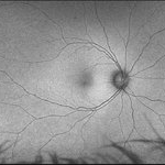
 Ocular Ischemia
Dec 12 2014 by David Callanan, MD
66-year-old white male, ocular ischemia.
Condition/keywords: ocular ischemic syndrome
 Loading…