Search results (4 results)
-
 Geographic Atrophy - Case 1: Photo 4 of 6
Oct 4 2012 by Gregg T. Kokame, MD, MMM, FASRS
NIRAF (near infrared autofluorescence) Image of patient with Geographic Atrophy
Photographer: Jaclyn Pisano, Retina Consultants of Hawaii
Imaging device: Heidelberg Spectralis
Condition/keywords: autofluorescence imaging, geographic atrophy, near infrared autofluorescence (NIRAF)
-
 Geographic Atrophy - Case 1: Photo 1 of 6
Oct 4 2012 by Gregg T. Kokame, MD, MMM, FASRS
NIRAF (near infrared autofluorescence) Image of patient with Geographic Atrophy
Photographer: Jaclyn Pisano, Retina Consultants of Hawaii
Imaging device: Heidelberg Spectralis
Condition/keywords: autofluorescence imaging, geographic atrophy, near infrared autofluorescence (NIRAF)
-
 Acute Macular Neuroretinopathy
Dec 11 2019 by Lauren Whaley
34-year-old female patient presented with changes in vision after recent upper respiratory infection. Referring doctor originally thought it was a blood pressure issue. She noticed a "C" shape in her vision. Infrared image was captured showing exactly what patient was describing! Doctor confirmed with this image that it was AMN.
Photographer: Lauren R. Whaley, COA
Imaging device: Heidelberg Spectralis
Condition/keywords: 30 degrees, acute macular neuroretinopathy, Heidelburg Spectralis, left eye, macula, near infrared autofluorescence (NIRAF)
-
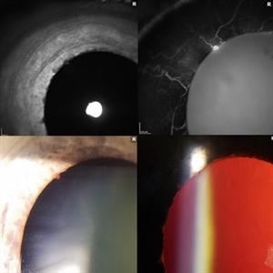 Iris Vascular Tuft
Jul 5 2022 by Olivia Rainey
Anterior segment imaging of a 66-year-old male with Vascular Disorders of Iris and Ciliary Body affecting his right eye. The physician stated that the findings are most consistent with a benign vascular tuft at the pupillary margin. The patient presented at the office with 20/20 vision in both eyes and had no ocular complaints at the time of his appointment.
Photographer: Olivia Rainey, OCT-C, COA
Imaging device: Heidelberg Spectralis, Slit Lamp with Samsung Galaxy 7
Condition/keywords: anterior segment, fluorescein angiogram (FA), heidelberg spectralis, infrared image, near infrared autofluorescence (NIRAF), slit lamp photo, vascular anomaly, vascular disorders of iris and ciliary body, vascular tuft
 Loading…