Search results (102 results)
-
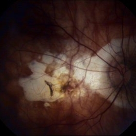
 Myopic Degeneration - Fundus Image
Oct 3 2013 by Gerardo Garcia-Aguirre, MD
Myopic degeneration - fundus image.
Condition/keywords: fundus photograph, myopic degeneration
-
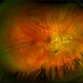
 Myopic macular degeneration
Jan 11 2013 by Alex P. Hunyor, MD
Myopic macular degeneration, left eye - extensive chorioretinal atrophy.
Condition/keywords: myopic degeneration, myopic fundus, myopic macular degeneration
-
 Myopic Degeneration
-
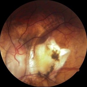
 Myopic Degeneration With RPE Loss
Oct 31 2013 by Jason S. Calhoun
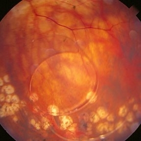
Patient in for follow up for glaucoma. VA is 20/30 in both eyes. Fundus photography shows the RPE loss due to myopic degeneration, nasally, inferiority in the left eye.
Photographer: Jason S. Calhoun, Ophthalmic Photographer, Department of Ophthalmology, Mayo Clinic Jacksonville
Imaging device: TOPCON TRC 50-EX
Condition/keywords: myopic degeneration
-
 Myopic Degeneration - Fundus Image
Oct 3 2013 by Gerardo Garcia-Aguirre, MD
Myopic degeneration - fundus image.
Condition/keywords: fundus photograph, myopic degeneration
-
 Myopic Degeneration - Fluorescein Angiography
Oct 3 2013 by Gerardo Garcia-Aguirre, MD
Myopic degeneration - fluorescein angiogram.
Condition/keywords: myopic degeneration
-
 Myopic Degeneration - Fluorescein Angiography
Oct 3 2013 by Gerardo Garcia-Aguirre, MD
Myopic degeneration - fluorescein angiogram.
Condition/keywords: myopic degeneration
-
 Myopic Degeneration - Fluorescein Angiography
Oct 3 2013 by Gerardo Garcia-Aguirre, MD
Myopic degeneration - fluorescein angiogram.
Condition/keywords: myopic degeneration
-
 Myopic Degeneration - Fluorescein Angiography
Oct 3 2013 by Gerardo Garcia-Aguirre, MD
Myopic degeneration - fluorescein angiogram.
Condition/keywords: myopic degeneration
-
 Myopic Degeneration - Fluorescein Angiography
Oct 3 2013 by Gerardo Garcia-Aguirre, MD
Myopic degeneration - fluorescein angiogram.
Condition/keywords: myopic degeneration
-
 Myopic Degeneration
Jul 3 2018 by Armando L. Oliver, MD
Myopic Degeneration
Photographer: Moises Castro
Imaging device: Optos California
Condition/keywords: pathologic myopia, posterior staphyloma
-
 LIO Dipped in the Vitreo
Aug 29 2016 by JEFFERSON R SOUSA, Tecg.º (Biomedical Systems Technology)
Patient Male, 51-years-old, with treatment with laser photocoagulation in myopic degeneration peripheral. Did FEC. suffered trauma (elbow) and had LIO dipped in the víteo.
Photographer: JEFFERSON R SOUSA - Institute Dr. Suel Abujamra / São Paulo - Brazil
Imaging device: Topcon TRC-50VT, Film, Kodak Ektachrome 160 - ASA 100 / 35mm, field of 35 degrees. Flash 100.
Condition/keywords: lens, myopic degeneration
-
 Myopic Degeneration - Red Free
Oct 3 2013 by Gerardo Garcia-Aguirre, MD
Myopic degeneration - red free.
Condition/keywords: myopic degeneration, red-free
-
 Myopic Degeneration, Macular Hemorrhage
Sep 10 2014 by Mehul A Shah
A 50-year-old male patient presented with complaint of sudden loss of vision.
Photographer: Drashti Netralaya,Dahod
Imaging device: FF 450
Condition/keywords: myopic degeneration
-
 Myopic degeneration - Red-Free
Oct 3 2013 by Gerardo Garcia-Aguirre, MD
Myopic degeneration - red free.
Condition/keywords: myopic degeneration, red-free
-
 Myopic Degeneration / CNVM
Sep 9 2014 by David Callanan, MD
85-year-old patient, myopic degeneration / CNVM.
Condition/keywords: choroidal neovascular membrane (CNVM), myopic degeneration
-
 High Myopia
Apr 2 2019 by Gary R. Cook, MD, FACS
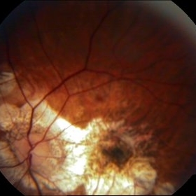
51-year-old white female with -7.00D myopia with a myopic conus on temporal aspect of the optic nerve and focal choroiretinal atrophy in the macula OS; V.A. = 20/25-1
Imaging device: Topcon VT-50
Condition/keywords: high myopia, myopic degeneration, myopic fundus, pathologic myopia
-
 Macular Hole
Jul 1 2014 by John S. King, MD
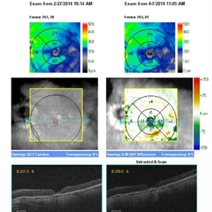
High myope c history of mac-off rrd c inf tear and ftmh that appeared chronic on presentation. RD repaired, hole remained open. ILMx performed with inverted flap. One and three months s/p MHx.
Photographer: Wayne A Ladlee Jr
Imaging device: Cirrus
Condition/keywords: macular hole, myopic degeneration
-
 Myopic Degeneration
Sep 10 2014 by Mehul A Shah
Chorio retinal atrophy.
Photographer: Drashti Netralaya
Imaging device: Zeiss FF450
Condition/keywords: myopia
-
 Myopic Degeneration
Jul 3 2018 by Armando L. Oliver, MD
Myopic Degeneration
Photographer: Moises Castro
Imaging device: Optos California
Condition/keywords: pathologic myopia, posterior staphyloma
-
 Prominent Long Ciliary Nerve
Jan 25 2022 by Kachelle Brown
Ultra-wide field photograph of a 48-year-old female with a prominent long ciliary nerve. Patient presented asymptomatic, and was referred for a macula on retinal detachment. Patient was diagnosed with high myopia and a posterior vitreous detachment, and the physician discussed increased risk of floaters, myopic degeneration and retinal detachment associated with high myopia. -24.00 prior to cataract surgery OU per patient.
Photographer: Kachelle Brown
Imaging device: Optos California
Condition/keywords: fundus photograph, high myopia, long ciliary nerve, optos, right eye, ultra-widefield image
-
 Myopic Degeneration
Oct 4 2014 by Mehul A Shah
A 40-year-old male presented with complaint of gradual diminished vision.
Photographer: Drashti Netralaya,Dahod
Imaging device: Zeiss ff450
Condition/keywords: posterior staphyloma
-
 Myopic Degeneration
Jul 3 2018 by Armando L. Oliver, MD
Late Views IVFA
Photographer: Moises Castro
Imaging device: Optos California
Condition/keywords: pathologic myopia, posterior staphyloma
-
 Macular Hemorrhage
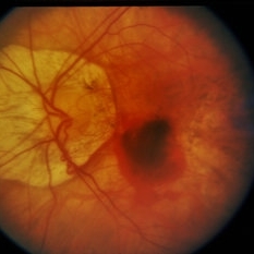
Apr 1 2019 by Gary R. Cook, MD, FACS
29-year-old white female with -12.25D high myopia OS and fresh macular hemorrhage; V.A.= 20/25.
Imaging device: Topcon VT-50
Condition/keywords: high myopia, lacquer cracks, macular hemorrhage, myopic degeneration
-
 Myopic Degeneration
Jul 3 2018 by Armando L. Oliver, MD
FAF
Photographer: Moises Castro
Imaging device: Optos California
Condition/keywords: pathologic myopia, posterior staphyloma
 Loading…