Search results (2 results)
-
 Slide 6-14
Feb 25 2019 by Lancaster Course in Ophthalmology
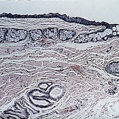
Dermoid cyst. The wall is lined by stratified squamous epithelium and contains epidermal appendages. The lumen, at top, contains keratin debris toward the left side (H&E x54).
Condition/keywords: cyst, lumen, stratified squamous epithelium
-
 Oval Pigmented Vitreous Cyst
Nov 27 2024 by Xinyu Zhao
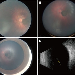
An 8-month-old infant was found to have a brown object in the left vitreous during a fundus screening. A wide-field digital retinal camera (RetCam) revealed a pigmented, non-transparent, freely floating, oval cystic lesion in the vitreous, measuring 2 disc diameters (Figures A-D). The cyst appeared cloudy when focused on the retina (Figure A) but was clearly defined in the vitreous (Figure B). Ultrasound showed a well-defined hyperreflective structure with a hyporeflective lumen (Figure D, indicated by the yellow arrow). A diagnosis of a vitreous pigment cyst, rare in infants, was made. Long-term follow-up is necessary to monitor changes affecting the infant’s vision.
Photographer: Xinyu Zhao, Shenzhen Eye Hospital, Shenzhen, China
Imaging device: RetCam
Condition/keywords: infant, vitreous cyst
 Loading…