Search results (7 results)
-
 Keratoconus
Oct 2 2013 by Jerald A. Bovino, MD
This is a profile picture of a patient with keratoconus.
Condition/keywords: cornea, degeneration, ectasia, thinning
-
 Keratoconus
Jul 11 2013 by Jason S. Calhoun
Side panel view of keratoconus of the cornea.
Photographer: Jason S. Calhoun, Department of Ophthalmology, Mayo Clinic Jacksonville, Florida
-
 Slide 4-24
Feb 20 2019 by Lancaster Course in Ophthalmology
Posterior keratoconus (Peter's anomaly). Peripheral cornea showing mesodermal dysgenesis ( x16).
Condition/keywords: cornea, dysgenesis, Keratosis pilaris (KP), Peters anomaly
-
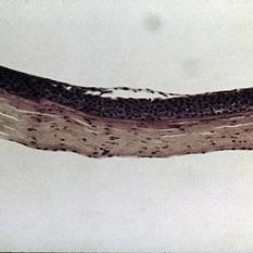 Slide 7-41
Feb 25 2019 by Lancaster Course in Ophthalmology
Keratoconus showing central thinning and scarring of the corneal button.
Condition/keywords: cornea, scar
-
 Keratoconus - Corneal Scar
May 3 2023 by Paula Lavigne
Keratoconus - Corneal Scar
Photographer: Paula Lavigne, Obras Sociais Irmã Dulce
Condition/keywords: corneal ectasia, corneal scars and opacities, keratoconus
-
 Enucleated Eye with Retinal Atrophy, Vitreomacular Traction and Keratoconus
May 18 2020 by McGill University Health Centre
This enucleation specimen shows areas of retinal atrophy (*) and areas of vitreomacular traction (arrow). This specimen also demonstrates keratoconus: a degenerative disorder of the eye in which the cornea thins and distorts into a pronounced conical shape (arrowhead). The keratoconus and vitreomacular tractions are unrelated.
Condition/keywords: atrophy, keratoconus, vitreomacular traction (VMT)
-
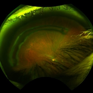 Corneal Ring Fundus Optos Photo
May 18 2020 by Catalina Montoya, MD
Temporal Fundus photograph right eye. Optos. No lesions in retina. Corneal Ring observed over fundus photograph. Patient with keratoconus.
Photographer: Catalina Montoya, Intermédica 1313. Medellín, Colombia
Imaging device: Optos
Condition/keywords: cornea
 Loading…