Search results (57 results)
-
 Cone-Rod Dystrophy
Mar 15 2017 by Hamid Ahmadieh, MD
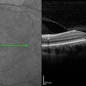
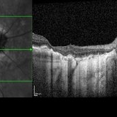
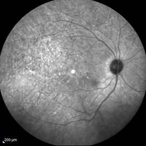
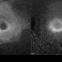
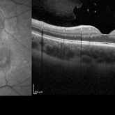
Infrared and OCT images of the left eye of a 16-year-old boy with decreased visual acuity and color vision deficiency due to cone-rod dystrophy.
Photographer: Abazarnezhad , Negah Eye Center, Tehran, Iran
Imaging device: Spectralis OCT
Condition/keywords: cone dystrophy, infrared image, optical coherence tomography (OCT)
-
 Choroidal Melanoma
Feb 2 2018 by Olivia Rainey
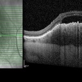
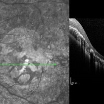
Optical coherence tomography with enhanced depth imaging of a 78-year-old female with choroidal melanoma with subretinal fluid affecting her right eye.
Photographer: Olivia Rainey
Imaging device: Heidelberg Spectralis
Condition/keywords: enhanced depth imaging, infrared image, optical coherence tomography (OCT), subretinal fluid, superior retina
-
 Geographic Atrophy - Case 1: Photo 3 of 6
Oct 4 2012 by Gregg T. Kokame, MD, MMM, FASRS
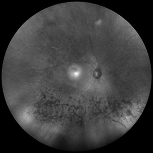
Infrared Image of patient with Geographic Atrophy
Photographer: Jaclyn Pisano, Retina Consultants of Hawaii
Imaging device: Heidelberg Spectralis
Condition/keywords: autofluorescence imaging, geographic atrophy
-
 "NVD Flower"
Oct 20 2023 by Daniel Davis, OCT-C
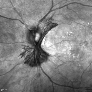
Infrared image of NVD (52F)
Imaging device: Heidelberg Spectralis
Condition/keywords: neovascularization of the disc (NVD)
-
 Best Disease
Mar 9 2013 by Hamid Ahmadieh, MD
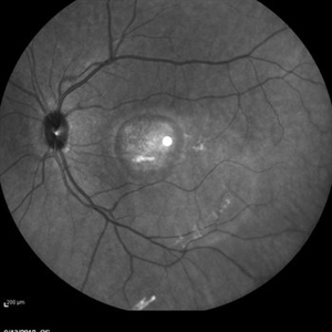
Infrared imaging of the left eye of a 49-year-old man with decreased VA due to advanced Best disease.
Photographer: Soodabeh Fooladin, Negah Eye Center, Tehran
Imaging device: Heidelberg Spectralis
Condition/keywords: Best disease, infrared image
-
 Proliferative Diabetic Retinopathy
Sep 15 2012 by Hamid Ahmadieh, MD
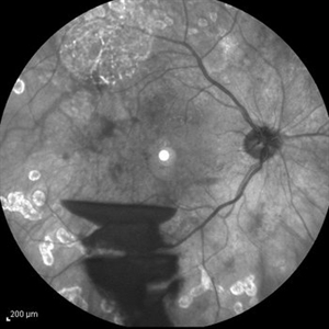
Infrared image of a 30-year-old woman with the history of scatter laser photocoagulation and a preretinal hemorrhage due to active PDR .
Photographer: Hamid Ahmadieh, MD, Ophthalmic Research Center, Labbafinejad Medical Center, Shahid Beheshti University of Medical Sciences
Imaging device: Heidelberg HRA
Condition/keywords: infrared image, preretinal hemorrhage
-
 Geographic Atrophy - Case 1: Photo 6 of 6
Oct 4 2012 by Gregg T. Kokame, MD, MMM, FASRS
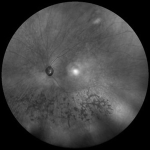
Infrared Image of patient with Geographic Atrophy
Photographer: Jaclyn Pisano, Retina Consultants of Hawaii
Imaging device: Heidelberg Spectralis
Condition/keywords: autofluorescence imaging, geographic atrophy
-
---thumb.jpg/image-square;max$300,300.ImageHandler) Polypoidal Choroidal Vasculopathy: Case 1 - Image 3 of 7
Polypoidal Choroidal Vasculopathy: Case 1 - Image 3 of 7
Oct 4 2012 by Gregg T. Kokame, MD, MMM, FASRS
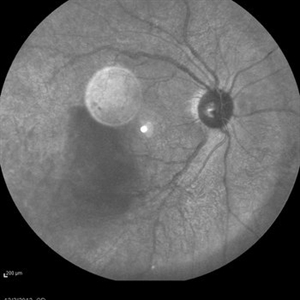
Fluorescein Angriography image of a 57-year-old woman with treatment-naive polypoidal choroidal vasculopathy. Series of images provides an comparative view of the same condition while utilizing a variet of different imaging procedures.
Photographer: Andrew Yuen, Retina Consultants of Hawaii
Imaging device: Heidelberg Spectralis
Condition/keywords: infrared image, polypoidal choroidal vasculopathy (PCV)
-
 Optic Nerve Head Drusen with OCT
Feb 2 2018 by Olivia Rainey
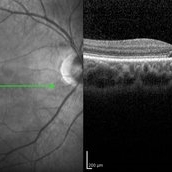
Optical coherence tomography with enhanced depth imaging of a 86-year-old male with optic nerve head drusen affecting his right eye. This patient has also been diagnosed with pseudoxanthoma elasticum and macular degeneration.
Photographer: Olivia Rainey
Imaging device: Heidelberg Spectralis
Condition/keywords: enhanced depth imaging, infrared image, macular degeneration, optic disc drusen, optic nerve, optical coherence tomography (OCT), pseudoxanthoma elasticum (PXE)
-
---thumb.jpg/image-square;max$300,300.ImageHandler) Primary Subhyaloid Hemorrhage Due to Valsalva Retinopathy
Primary Subhyaloid Hemorrhage Due to Valsalva Retinopathy
Nov 13 2013 by Hamid Ahmadieh, MD
Infrared image of the left eye of a 25-year-old man with primary subhyaloid hemorrhage due to Valsalva retinopathy.
Photographer: Nayereh Hadipour, Negah Eye Center, Tehran
Imaging device: Heidelberg Spectralis
Condition/keywords: infrared image, subhyaloid hemorrhage, valsalva retinopathy
-
 Silicone Oil Bubble in Anterior Chamber - 30 Degree Angle
Apr 11 2016 by Zach Dupureur
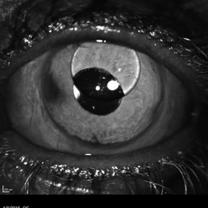
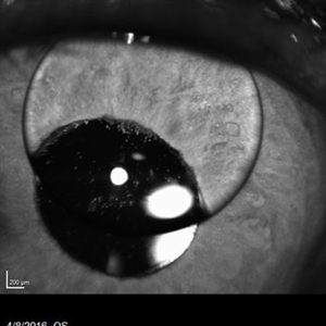
30 % silicone oil bubble involving central visual axis. Occurred after a PPV with silicone oil. Oil from vitreous moved into the anterior chamber.
Photographer: Zachary Dupureur, OCT-C
Imaging device: Heidelberg Spectralis
Condition/keywords: anterior chamber, detachment, infrared image, pars plana vitrectomy (PPV), scleral buckle, silicone oil
-
 Case 2 Retinitis Pigmentosa BAF IRAF OD
May 14 2014 by Avris Romario Diparaja Siahaan
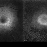
Fundus image a 57-year-old man with retinitis pigmentosa on both eyes. These image were taken with blue auto fluorescein mode (BAF) and infrared auto fluorescence (IRAF).
Photographer: Avris Romario Diparaja Siahaan
Imaging device: Heidelberg HRA + OCT Spectralis
Condition/keywords: autofluorescence imaging, fundus photograph, infrared image, retinitis pigmentosa
-
 Fundus Flavimaculatus and CNV
Nov 14 2013 by Hamid Ahmadieh, MD
Infrared image of the right eye of a 35-year-old woman with subfoveal CNV secondary to fundus flavimaculatus .
Photographer: Nayereh Hadipour, Negah Eye Center, Tehran
Condition/keywords: choroidal neovascularization (CNV), fundus flavimaculatus, infrared image, retinal flecks
-
 Stargardts Disease
Mar 13 2013 by Hamid Ahmadieh, MD
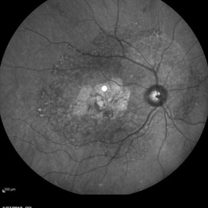
Infrared image of the right eye of a 46-year-old man with decreased VA due to advanced Stargardts disease.
Photographer: Nayereh Hadipoor, Negah Eye Center, Tehran
Imaging device: Heidelberg Spectralis
Condition/keywords: infrared image, Stargardt disease
-
 Cone-Rod Dystrophy
Mar 15 2017 by Hamid Ahmadieh, MD
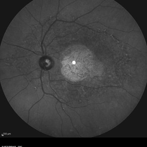
Infrared and OCT images of the right eye of a 16-year-old boy with decreased visual acuity and color vision deficiency due to cone-rod dystrophy.
Photographer: Abazarnezhad , Negah Eye Center, Tehran, Iran
Imaging device: Spectralis OCT
Condition/keywords: cone dystrophy, infrared image, optical coherence tomography (OCT)
-
 Stargardt's Disease
Mar 13 2013 by Hamid Ahmadieh, MD
Infrared image of the right eye of a 46-year-old man with decreased VA due to advanced Stargardt's disease.
Photographer: Nayereh Hadipoor, Negah Eye Center, Tehran
Imaging device: Heidelberg Spectralis
Condition/keywords: infrared image, Stargardt disease
-
 Case 2 Retinitis Pigmentosa BAF IRAF OS
May 14 2014 by Avris Romario Diparaja Siahaan
Fundus image a 57-year-old man with retinitis pigmentosa on both eyes. These image were taken with blue auto fluorescein mode (BAF) and infrared auto fluorescence (IRAF).
Photographer: Avris Romario Diparaja Siahaan
Imaging device: Heidelberg HRA + OCT Spectralis
Condition/keywords: autofluorescence imaging, fundus photograph, infrared image, retinitis pigmentosa
-
 Sector Retinitis Pigmentosa
Mar 13 2014 by Hyung-Woo Kwak, MD
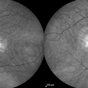
Wide field infrared image of an 57-year-old woman with a sector retinitis pigmentosa. Regionalized areas of bone spicule pigmentation is in the inferior quadrants of the retina.
Photographer: Missok Lee, Kyung Hee University Hospital, Seoul, Korea
Imaging device: Heidelberg Spectralis
Condition/keywords: sector retinitis pigmentosa
-
 Sector Retinitis Pigmentosa
Mar 13 2014 by Hyung-Woo Kwak, MD
Wide field infrared image of an 57-year-old woman with a sector retinitis pigmentosa. Regionalized areas of bone spicule pigmentation is in the inferior quadrants of the retina.
Photographer: Missok Lee, Kyung Hee University Hospital, Seoul, Korea
Imaging device: Heidelberg Spectralis
Condition/keywords: sector retinitis pigmentosa
-
 Behcet's Disease
Mar 13 2013 by Hamid Ahmadieh, MD
Infrared image of the right eye of a 23-year-old man with retinal vasculitis and branch retinal vein occlusion (BRVO) due to Behcet's disease .
Photographer: Solmaz Shahmohammad, Negah Eye Center, Tehran
Imaging device: Heidelberg Spectralis
Condition/keywords: branch retinal vein occlusion (BRVO), infrared image, retinal vasculitis
-
 Hypotony
May 29 2013 by Zofia Anna Nawrocka (vel Michalewska), MD, PhD
Infrared image of a 75-year-old patient with hypotony, 2 weeks after trauma, 2 years after extracapsular cataract surgery.
Photographer: Zofia Michalewska, Ophthalmic Clinic "Jasne Blonia
Imaging device: Spectralis
Condition/keywords: hypotony
-
 Silicone Oil Bubble in Anterior Chamber - 20 Degree Angle
Apr 11 2016 by Zach Dupureur
30 % silicone oil bubble involving central visual axis. Occurred after a PPV with silicone oil. Oil from vitreous moved into the anterior chamber.
Photographer: Zachary Dupureur, OCT-C
Imaging device: Heidelberg Spectralis
Condition/keywords: anterior chamber, detachment, infrared image, pars plana vitrectomy (PPV), scleral buckle, silicone oil
-
 Multiple Astrocytic Hamartomas
Jul 26 2018 by Olivia Rainey
Optical coherence tomography of a 7-year-old female with multiple astrocytic harmartomas as a retinal manifestation of tuberous sclerosis. Patient came to our office to rule out possible drug toxicity from Sabril, a an anticonvulsant. There were no signs of retinal toxicity by extended ophthalmoscopy or imaging, yet she will be monitored every 6 months.
Photographer: Olivia Rainey
Imaging device: Heidelberg Spectralis
Condition/keywords: astrocytic hamartoma, Heidelburg Spectralis, infrared image, left eye, optical coherence tomography (OCT), tuberous sclerosis
-
 Macular Coloboma and Pigmentary Retinopathy
Feb 25 2017 by Hamid Ahmadieh, MD
Infrared and OCT images of the left eye of a 25-year-old woman with bilateral macular colobomata and pigmentary retinopathy similar to Leber's congenital amaurosis.
Photographer: Shabnam Poureh, Negah Eye Center, Tehran, Iran
Condition/keywords: bilateral pigmentary retinopathy, infrared image, macular coloboma, optical coherence tomography (OCT)
-
 Central Areolar Choroidal Dystrophy
Jul 7 2015 by Hamid Ahmadieh, MD
Infrared image of both eyes of a 58-year-old man with progressive loss of vision. VA OD is 20/60 and VA OS is 20/400.
Photographer: Soulmaz Shahmohammad, Negah Eye Center, Tehran, Iran
Imaging device: Specteralis
Condition/keywords: central areolar choroidal dystrophy (CACD), infrared image
 Loading…