Search results (42 results)
-
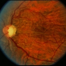
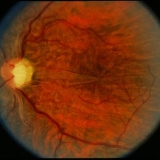
 Choroidal folds due to hypotony
Jan 11 2013 by Alex P. Hunyor, MD
Choroidal folds due to hypotony
Condition/keywords: choroidal folds, hypotonous retinopathy
-
 Uveitic Hypotony
Dec 10 2012 by Yale L. Fisher, MD
Hypotony can produce specific findings on ultrasound examination such as diffuse thickening of the choroid, scleral thickening, and shortened axial length.
Condition/keywords: choroid, hypotony, scleral thickening, uveitis, video
-
 Ocular Hypotony Due to Leaking Bleb
Apr 1 2019 by Anfisa Ayalon, MD
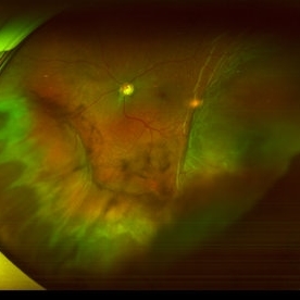
81-year-old male who had trabeculectomy in his right eye 4 years ago, presented to the emergency room with complains of decreased vision in that eye for two months. Slit-lamp examination showed cystic bleb with leakage, intraocular pressure was 0 MMHg. Fundus examination showed hypotony maculopathy, peripheral choroidal detachments, multiple chorioretinal folds with subretinal fluid.
Photographer: Anfisa Ayalon, MD., Meir Medical Center, Kfar Saba, Israel.
Imaging device: California, Optos 200 DTX
Condition/keywords: choroidal detachment, hypotonous retinopathy, hypotony maculopathy
-
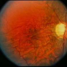
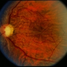
 PRE CF OD June 5, 2013
Mar 12 2014 by Manish Nagpal, MD, FRCS (UK), FASRS
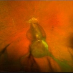
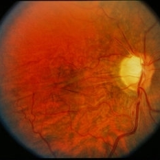
Fundus photo of a 32-year-old male presenting with post traumatic choroidal folds and hypotony.
Photographer: Pooja Barot
Condition/keywords: choroidal folds
-
 SD-OCT of Ocular Hypotony
May 29 2013 by Zofia Anna Nawrocka (vel Michalewska), MD, PhD
SD-OCT of a 75-year-old patient with hypotony, 2 weeks after trauma, 2 years after extracapsular cataract surgery.
Photographer: Zofia Michalewska, Ophthalmic Clinic "Jasne Blonia
Imaging device: Spectralis
Condition/keywords: hypotony, optical coherence tomography (OCT)
-
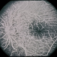
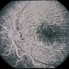
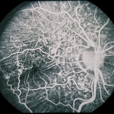
 Fluorescein Angiography of Ocular Hypotony
May 29 2013 by Zofia Anna Nawrocka (vel Michalewska), MD, PhD
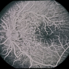
Fluorescein angiography of a 7- year-old patient with hypotony, 2 weeks after trauma, 2 years after extracapsular cataract surgery.
Photographer: Zofia Michalewska, Ophthalmic Clinic "Jasne Blonia
Imaging device: Spectralis
Condition/keywords: hypotony
-
 Autofluorescence of Ocular Hypotony
May 29 2013 by Zofia Anna Nawrocka (vel Michalewska), MD, PhD
Autofluorescence image of a 75-year-old patient with hypotony, 2 weeks after trauma, 2 years after extracapsular cataract surgery.
Photographer: Zofia Michalewska, Ophthalmic Clinic "Jasne Blonia
Imaging device: Spectralis
Condition/keywords: hypotony
-
 Suprachoroidal Hemorrhage
Sep 2 2020 by Rinal Pandit
Fundus photograph of left eye of a 56-year-old female with primary angle closure glaucoma showing massive hemorrhagic choroidal detachment that developed following trabeculectomy surgery. Suprachoroidal hemorrhage is defined as the accumulation of blood within the potential space between the choroid and sclera, with the source of the blood being the long or short posterior ciliary artery. Delayed suprachoroidal hemorrhage (DSHC) remains one of the most dreaded and sight threatening complications of glaucoma filtration surgery. The risk factors include old age, hypertension, high myopia, arteriosclerosis, chronically elevated IOP, sudden hypotony, trauma, aphakia/pseudophakia, prior vitrectomy, history of 5 FU injections and anti-platelet agents. The incidence of postoperative SCH after trabeculectomy varies between 0.6%- 1.4%. DSCH after surgery varies considerably in severity but is generally characterized by the sudden onset of severe pain, decreased vision, and a shallow anterior chamber usually associated with raised intraocular pressure. B-scan ultrasonography can help to distinguish serous from hemorrhagic choroidals.Suprachoroidal hemorrhages appear as dome-shaped elevations of the retina with increased echo densities that are often heterogeneous and within the suprachoroidal space. Choroidal effusions appear as dome-shaped elevations with hypoechoic suprachoroidal space. The first step in the management is the timely diagnosis. Medical management includes oral and topical antiglaucoma drugs to lower IOP, oral and topical steroids to control inflammation and topical cycloplegics and oral analgesics to tackle pain. Serial ultrasound B scans of the affected eye should be performed in order to monitor progression of the SCH and help determine apposition, height, and liquefaction of the SCH. Indications of surgical drainage include non resolution with medical management,concurrent retinal detachment, central retinal apposition (kissing choroidals) and incarceration of vitreous in the wound site. The ideal time of drainage is between 7-14 days depending upon clot lysis. The prognosis of both intraoperative and postoperative SCH is poor. An overwhelming majority of patients do not achieve pre-hemorrhage visual acuity and most do not recover to a visual acuity of 20/200 or better. The major determinants of good or bad visual outcomes of SCH’s are preoperative visual acuity and retinal detachment at the time of hemorrhage, respectively.
Imaging device: OPTOS,Ultra wide field retinal imaging system
Condition/keywords: suprachoroidal hemorrhage, trabeculectomy, ultra-wide field imaging
-
 Hypotony
May 29 2013 by Zofia Anna Nawrocka (vel Michalewska), MD, PhD
Infrared image of a 75-year-old patient with hypotony, 2 weeks after trauma, 2 years after extracapsular cataract surgery.
Photographer: Zofia Michalewska, Ophthalmic Clinic "Jasne Blonia
Imaging device: Spectralis
Condition/keywords: hypotony
-
 Closed Funnel Retinal Detachment
Oct 8 2019 by Olivia Rainey
Ultra-wide field pseudocolor image of a 57-year-old male with a closed funnel retinal detachment with anterior and posterior napkin rings affecting his left eye. Patient presented with klebsiella endophthalmitis in UK, and was in medically induced coma with tracheostomy. He awoke after sedation with loss of vision in both eyes, later developing a retinal detachment in both eyes. Prior inflammation attributable to prephthisical state and chronic funnel retinal detachment. The eye is inoperable and observation is recommended.
Photographer: Olivia Rainey and Amber Poss
Imaging device: Optos
Condition/keywords: blind eye, funnel, hypotony, klebsiella endopthalmitis, left eye, Optos
-
 Hypotony from Over-Filtration
Mar 5 2014 by David Callanan, MD
45-year-old Hispanic male with hypotony from over-filtration.
Condition/keywords: hypotony
-
 Hypotony Maculopathy
Apr 1 2019 by Anfisa Ayalon, MD
Fundus autofluorescence image of 81-year-old male with right eye ocular hypotony due to leaking bleb. Note severe hypotony maculopathy, peripheral choroidal detachments, multiple chorioretinal folds.
Photographer: Anfisa Ayalon, MD., Meir Medical Center, Kfar Saba, Israel.
Imaging device: California, Optos 200 DTX
Condition/keywords: choroidal detachment, choroidal folds, fundus autofluorescence (FAF), hypotonous retinopathy, hypotony maculopathy
-
 Hypotony Maculopathy
May 3 2018 by Alexandr Stepanov
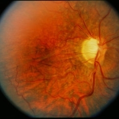
Hypotony maculopathy.
Photographer: Alexandr Stepanov MD, PhD, FEBO, Faculty Hospital Hradec Kralove, Czech Republic
Condition/keywords: hypotony maculopathy
-
 Hypotony from Over-Filtration
Mar 5 2014 by David Callanan, MD
45-year-old Hispanic male with hypotony from over-filtration.
Condition/keywords: hypotony
-
 Hypotony from Over-Filtration
Mar 5 2014 by David Callanan, MD
45-year-old Hispanic male with hypotony from over-filtration.
Condition/keywords: hypotony
-
 Hypotony from Over-Filtration
Mar 5 2014 by David Callanan, MD
45-year-old Hispanic male with hypotony from over-filtration.
Condition/keywords: hypotony
-
 Hypotony from Over-Filtration
Mar 5 2014 by David Callanan, MD
45-year-old Hispanic male with hypotony from over-filtration.
Condition/keywords: hypotony
-
 Hypotony from Over-Filtration
Mar 5 2014 by David Callanan, MD
45-year-old Hispanic male with hypotony from over-filtration.
Condition/keywords: hypotony
-
 Hypotony from Over-Filtration
Mar 5 2014 by David Callanan, MD
45-year-old Hispanic male with hypotony from over-filtration.
Condition/keywords: hypotony
-
 Hypotony from Over-Filtration
Mar 5 2014 by David Callanan, MD
45-year-old Hispanic male with hypotony from over-filtration.
Condition/keywords: hypotony
-
 Traumatic Giant Retinal Tear Associated Retinal Detachment
Nov 9 2019 by Luis J Haddock, MD
This wide field fundus photograph of the left eye shows a traumatic giant retinal tear associated with total retinal detachment. The image shows the torn superior retina folded over the macula with the underside of the retina visible. There is associated peripheral choroidal detachment due to hypotony from giant retinal tear. This patient has history of spondyloepithelial dysplasia with dwarfism and presented with vision loss after a recent blunt trauma with elbow to the eye.
Imaging device: Optos
Condition/keywords: giant retinal tear, traumatic optic neuropathy
-
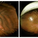 Scleral Infolding Due to Hypotony
Feb 17 2015 by Danielle Strauss, MD FASRS
Wide field imaging of the left eye in a patient with scleral infolding due to hypotony from ruptured trabeculectomy. Left side image shows the eye prior to revision of the trabeculectomy and pars plana vitrectomy. Right side shows post-op image of the eye.
Photographer: Robert Masini, New York Eye and Ear Infirmary of Mount Sinai
Condition/keywords: hypotony
-
 Hypotony, Thickend Choroid, Total Retinal Detachment, Foreshortened Globe
Dec 10 2012 by Yale L. Fisher, MD
Foreshortening in a phtisical eye with a thickened choroid.
Condition/keywords: video
-
 Hypotony from Over-Filtration
Mar 5 2014 by David Callanan, MD
45-year-old Hispanic male with hypotony from over-filtration.
Condition/keywords: hypotony
-
 Hypotony from Over-Filtration
Mar 5 2014 by David Callanan, MD
45-year-old Hispanic male with hypotony from over-filtration.
Condition/keywords: hypotony
 Loading…