Search results (37 results)
-
 Advanced PDR
Sep 29 2012 by Hamid Ahmadieh, MD
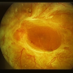
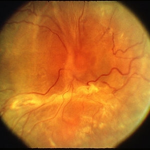
Color fundus photograph of a 32-year-old man with insulin-dependent diabetes mellitus and regressed advanced PDR with severe fibrous proliferation and traction retinal detachment sparing the macula.
Photographer: Hamid Ahmadieh, MD; Ophthalmic Research Center, Labbafinejad Medical Center, Shahid Beheshti University of Medical Sciences
Condition/keywords: fibrous proliferation, tractional retinal detachment
-
 Advanced PDR
Sep 29 2012 by Hamid Ahmadieh, MD
Color fundus photograph of a 32-year-old man with insulin-dependent diabetes mellitus and regressed advanced PDR with severe fibrous proliferation and traction retinal detachment sparing the macula.
Photographer: Hamid Ahmadieh, MD; Ophthalmic Research Center, Labbafinejad Medical Center, Shahid Beheshti University of Medical Sciences
Condition/keywords: fibrous proliferation, tractional retinal detachment
-
---thumb.jpg/image-square;max$300,300.ImageHandler) Fibrovascular Proliferation
Fibrovascular Proliferation
Feb 13 2013 by From the Collections of Thomas M. Aaberg, MD and Thomas M. Aaberg Jr., MD
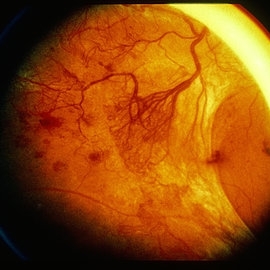
Neovascularization, fibrous proliferation, intraretinal hemorrhage.
Condition/keywords: fibrous proliferation, intraretinal hemorrhage, neovascularization (NV)
-
 Severe fibrovascular proliferative from PDR
Jan 1 2013 by John T. Thompson, MD
Severe fibrovascularization proliferation in eye with PDR.
Condition/keywords: fibrous proliferation, retinal neovascularization
-
 NVD with fibrosis from PDR
Jan 1 2013 by John T. Thompson, MD
Fibrovascular proliferation from PDR, florid NVD.
Condition/keywords: fibrous proliferation, neovascularization of the disc (NVD)
-
 Severe Fibrovascular Proliferation From PDR
Jan 1 2013 by John T. Thompson, MD
Severe fibrovascular proliferation associated with PDR.
Condition/keywords: fibrous proliferation
-
 Advanced Active PDR
Mar 29 2013 by Henry J. Kaplan, MD
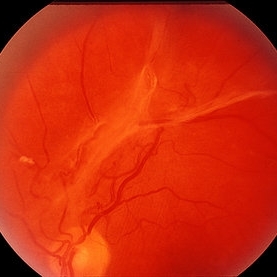
Large active NVEs with fibrous proliferations in diabetes.
Condition/keywords: fibrous proliferation, neovascularization (NV)
-
 PDR with fibrous proliferation
Jan 1 2013 by John T. Thompson, MD
Fibrous proliferation associated with PDR.
Condition/keywords: fibrous proliferation
-
 PDR
PDR
Mar 29 2013 by Henry J. Kaplan, MD
Fibrous proliferation (FPE) in a patient with PDR.
Condition/keywords: fibrous proliferation, FPE
-
 Advanced Proliferative Diabetic Retinopathy
Nov 4 2017 by Hamid Ahmadieh, MD
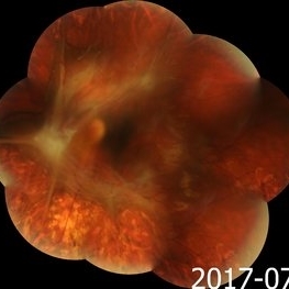
Merged color fundus photograph of the left eye of a 30-year-old woman with type1 diabetes since childhood. Note laser scars, severe fibrous proliferation, traction RD and macular dragging.
Photographer: Shabnam Poureh, Negah Eye Center, Tehran, Iran
Condition/keywords: color fundus photograph, diabetes, fibrous proliferation, proliferative diabetic retinopathy (PDR), severe traction
-
 Fibrotic Tractional Membrane in ROP Stage 5
Nov 7 2013 by Maria Ana Martinez-Castellanos, MD
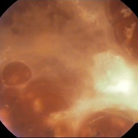
Stage 5 retinopathy of prematurity in a 6 month old baby.
Photographer: Maria A. Martinez-Castellanos. Asociacion para Evitar la Ceguera en Mexico
Imaging device: RetCam II
Condition/keywords: fibrous proliferation, fibrovascular proliferation, retinopathy of prematurity (ROP)
-
 TRD
TRD
Mar 29 2013 by Henry J. Kaplan, MD
Advanced fibrous proliferation and TRD in diabetes.
Condition/keywords: tractional retinal detachment
-
 Fibrovascular Membrane
Apr 5 2018 by Mohamed Tawfik, MD
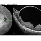
16 mm wide field OCT scan of a case of fiber-vascular membrane demonstrate the point of attachment of membrane.
Photographer: Mohamed A,Tawfik MD,FRCSed
Condition/keywords: fibrotic neovascularization, fibrous proliferation, fibrovascular change
-
 Proliferative Diabetic Retinopathy
Mar 25 2013 by Ratimir Lazic, MD, PhD
Color fundus photography of a 62- year-old diabetic patient. Severe fibrous proliferations with traction retinal detachment can be seen. Pars plana vitrectomy was preformed on that eye.
Photographer: Marko Lukic, MD
Imaging device: Zeis Visucam Lite 2
Condition/keywords: neovascularization (NV)
-
---thumb.jpg/image-square;max$300,300.ImageHandler) Proliferative Diabetic Retinopathy
Proliferative Diabetic Retinopathy
Mar 25 2013 by Ratimir Lazic, MD, PhD
Color fundus photography of a 62- year-old diabetic patient. Fibrous proliferations along upper temporal branch and posterior pole with no traction on retina can be seen. Suspected neovascularization nasal from PNO.
Photographer: Marko Lukic, MD
Imaging device: Zeis Visucam Lite 2
Condition/keywords: neovascularization (NV)
-
 Severe Fibrovascular Proliferative Mass With Tractional RD in PDR
Aug 1 2017 by Eitae Kim, MD
Severe fibrovascular proliferation with tractional retinal detachment is seen on UWF fundus photograph.
Photographer: Eitae Kim, BOIM retinal center, Pureun eye hospital
Condition/keywords: fibrous proliferation, tractional retinal detachment
-
---thumb.jpg/image-square;max$300,300.ImageHandler) Reduced Vision
Reduced Vision
Feb 4 2014 by Maurice F. Rabb
69-year-old female with a detachment of the pigment epithelium in the right eye with hemorrhagic and early fibrous proliferative changes. The left eye contained a large, turbid detachment of the pigment epithelium with patchy atrophy, a very shallow evident overlying sensory retinal detachment, but no subretinal hemorrhage. The visual acuity in each eye was 20/400.
Condition/keywords: fibrous proliferation, patchy atrophy, pigment epithelial detachment (PED), reduced vision
-
 Severe Fibrovascular Proliferative Mass With Tractional RD in PDR
Aug 1 2017 by Eitae Kim, MD
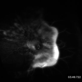
Severe fibrovascular proliferation with tractional retinal detachment is seen on UWF FAG.
Photographer: Eitae Kim, BOIM retinal center, Pureun eye hospital
Condition/keywords: fibrous proliferation, tractional retinal detachment
-
---thumb.jpg/image-square;max$300,300.ImageHandler) Reduced Vision
Reduced Vision
Feb 4 2014 by Maurice F. Rabb
69-year-old female with a detachment of the pigment epithelium in the right eye with hemorrhagic and early fibrous proliferative changes. The left eye contained a large, turbid detachment of the pigment epithelium with patchy atrophy, a very shallow evident overlying sensory retinal detachment, but no subretinal hemorrhage. The visual acuity in each eye was 20/400.
Condition/keywords: fibrous proliferation, patchy atrophy, pigment epithelial detachment (PED), reduced vision
-
---thumb.jpg/image-square;max$300,300.ImageHandler) Reduced Vision
Reduced Vision
Feb 4 2014 by Maurice F. Rabb
69-year-old female with a detachment of the pigment epithelium in the right eye with hemorrhagic and early fibrous proliferative changes. The left eye contained a large, turbid detachment of the pigment epithelium with patchy atrophy, a very shallow evident overlying sensory retinal detachment, but no subretinal hemorrhage. The visual acuity in each eye was 20/400.
Condition/keywords: fibrous proliferation, patchy atrophy, pigment epithelial detachment (PED), reduced vision
-
---thumb.jpg/image-square;max$300,300.ImageHandler) Reduced Vision
Reduced Vision
Feb 4 2014 by Maurice F. Rabb
69-year-old female with a detachment of the pigment epithelium in the right eye with hemorrhagic and early fibrous proliferative changes. The left eye contained a large, turbid detachment of the pigment epithelium with patchy atrophy, a very shallow evident overlying sensory retinal detachment, but no subretinal hemorrhage. The visual acuity in each eye was 20/400.
Condition/keywords: fibrous proliferation, patchy atrophy, pigment epithelial detachment (PED), reduced vision
-
 Ultra-Widefield Image of Tractional-Rhegmatogenous Retinal Detachment Sparing Fovea
Jul 16 2021 by Kushal S Delhiwala, MBBS, MS, FMRF,FICO, FAICO
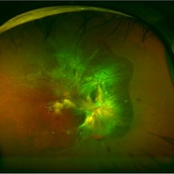
Ultra-widefield fundus photograph of an 45-year-old phakic male with superior tractional-rhegmatogenous retinal detachment sparing fovea. Retinal break was observed at the base of fibrous proliferation. Scattered whitish outer retinal spots were noted in area of retinal detachment.
Photographer: Kushal Delhiwala, Netralaya superspeciality eye hospital, Ahmedabad, Gujarat,India
Imaging device: Optos Daytona
Condition/keywords: fibrovascular proliferation, fibrovascular tissue, outer retinal white spots, tractional retinal detachment, ultra-wide field imaging
 Loading…