Search results (16 results)
-
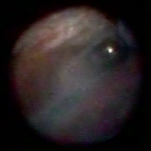
 Endoscopy: Giant Retinal Tear—Anterior Flap Incarceration
Dec 10 2012 by Yale L. Fisher, MD
This video demonstrates endoscopic visualisation of incarceration of the anterior flap of a giant retinal tear
Condition/keywords: endoscopy, retinal tear, video
-
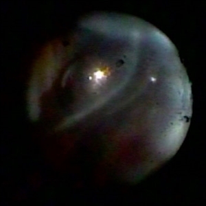
 Endoscopy: Infusion Cannula Positions
Dec 11 2012 by Yale L. Fisher, MD
Infusion cannula positions can be visualized with the endoscope, especially useful when microscopic views are limited or impossible as in this short movie. Here, the infusion cannula had been placed in a standard fashion but endoscopic imaging reveals partial engagement of peripheral retinal tissue. Manipulation of the tip under endoscopic viewing eliminates the undesired engagement and reveals the proper infusion position with no obstruction.
Condition/keywords: endoscopy, infusion cannula positions, video
-
 Endoscopy: Peripheral Endoscopic Ciliary Body Ablation for Control of Neovascular Glaucoma in Diabetic Patient
Dec 10 2012 by Yale L. Fisher, MD
With this patient microscopic control was not possible due to a small fixed pupil with vascularized synechiae to a posterior chamber IOL. There was recurrent intraocular bleeding and elevated IOP. The ciliary body ablation was accomplished first with a 532 laser fed through the working channel of the endoscope. Examination of the peripheral retina revealed a ring-like rhegmatogenous retinal separation and a large inferior tear with persistent traction. Endoscopically controlled imaging and a second instrument (suction/cutter) removed the tractional elements and permitted an air fluid exchange. Retinal reattachment occurred as the air-fluid exchange was completed permitting laser ablation of the ring like area that had been separated. The entire procedure was performed utilizing the small gauge endoscope.
Condition/keywords: video
-
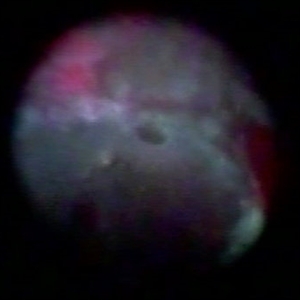
 Endoscopy: Open Angle Glaucoma and Retinal Detachment PVR
Dec 10 2012 by Yale L. Fisher, MD
Endoscopy: open angle glaucoma and retinal detachment PVR.
Condition/keywords: video
-
 Endoscopy: Shielded Endoscopic Cleaner
Dec 10 2012 by Yale L. Fisher, MD
Cleaning of the endoscope is possible intraocularly with a protected and extendible sponge like wiper (pat). The device is useful to remove water droplets and non-adherent debris, especially following an air fluid exchange with the endoscope located in the air bubble.
Condition/keywords: video
-
 Endoscopy: Ciliary Body
Dec 10 2012 by Yale L. Fisher, MD
Ciliary body imaging is possible with the endoscope when patients are aphakic or pseudophakic. The ciliary processes are easily visualized in this short movie clip.
Condition/keywords: video
-
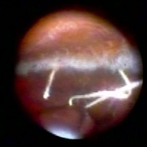
 Endoscopy: Open Angle Glaucoma and Recurrent Retinal Detachment
Dec 10 2012 by Yale L. Fisher, MD
Endoscopically controlled surgical repair of a rhegmatogenous retinal detachment in a phakic glaucoma patient with a fixed miotic pupil. The formed vitreous is still attached in the posterior pole but separated peripherally. A horseshoe tear is located in the superior nasal portion of the globe. The flap is trimmed and the posterior formed vitreous face is removed from the optic nerve and posterior pole with an open vertical scissors blade. An air fluid exchange through the break completes the reattachment.
Condition/keywords: video
-
 Endoscopy: Peripheral Lens Fragment
Dec 10 2012 by Yale L. Fisher, MD
Peripheral endoscopic imaging of the pars plana region. The second instrument can be seen in the sclerotomy site moving in and out. Scanning of the more anterior region reveals a large particle of retained lens material following phacoemulsification. This particle was responsible for recurrent inflammation and cystoid macular edema. Subsequent removal ended the inflammation and retinal edema.
Condition/keywords: video
-
 Endoscopy: Anterior Neovascularization- Diabetic Retinopathy I
Dec 10 2012 by Yale L. Fisher, MD
Another movie from Dr. Yale Fisher's collection of endoscopic imaging- this is another case of anterior neovascularization with diabetic retinopathy. No retinal detachment is present.
Condition/keywords: video
-
 Endoscopy: Anterior Neovascularization- Diabetic Retinopathy II
Dec 10 2012 by Yale L. Fisher, MD
Another movie from Dr. Yale Fisher's collection of endoscopic imaging- in this case, anterior neovascularization in a case of diabetic retinopathy. No retinal detachment is present.
Condition/keywords: video
-
 Endoscopy: An Introduction to Ophthalmic Endoscopy in Vitreoretinal Surgery
Dec 10 2012 by Yale L. Fisher, MD
Dr. Yale Fisher discusses the history, technique, and other basics of ophthalmic endoscopy for use in vitreoretinal surgery.
Condition/keywords: video
-
 Introduction to Ophthalmic Endoscopy
Dec 10 2012 by Yale L. Fisher, MD
Dr. Yale Fisher describes his approach to learning ophthalmic endoscopy.
Condition/keywords: video
-
 Endoscopic View: the Beginning of Anterior Hyaloid Membrane Pneumatic Dissection
Oct 2 2019 by Radwan S. Ajlan, MBBCh, FRCS(C)
Endoscopic view: the beginning of anterior hyaloid membrane pneumatic dissection.
Condition/keywords: endoscopy, hyaloid, membranes
-
 Endoscopic View - The Start of Anterior Hyaloid Membrane Pneumatic Dissection
Oct 2 2019 by Radwan S. Ajlan, MBBCh, FRCS(C)
Endoscopic view - the start of anterior hyaloid membrane pneumatic dissection.
Condition/keywords: endoscopy, hyaloid, membranes
-
 Endoscopic View of Anterior Proliferative Vitreoretinopathy, Retinal Tear, and Tractional Retinal Detachment
Oct 2 2019 by Radwan S. Ajlan, MBBCh, FRCS(C)
Endoscopic view of anterior proliferative vitreoretinopathy, retinal tear, and tractional retinal detachment.
Condition/keywords: endoscopy, proliferative vitreoretinopathy (PVR), tractional retinal detachment
-
 Endoscopic View of a Rotated Sclera Fixated Lens Implant
Oct 2 2019 by Radwan S. Ajlan, MBBCh, FRCS(C)
Endoscopic view of a rotated sclera fixated lens implant.
Condition/keywords: endoscopy, sclera
 Loading…