Search results (6 results)
-
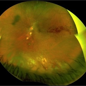
 Candy Stripe Sign
Mar 30 2023 by pedro fernandes souza neto
Candy Stripe Sign, patient with proliferative diabetic retinopathy progressing to vitreous hemorrhage and subsequently to ghost cell glaucoma.
Photographer: Marlos Henrique Oliveira Junior, Federal University of Bahia.
Condition/keywords: dehemoglobinized hemorrhage, diabetes, diabetic glaucoma
-
 Penetrating Trauma of an Inadvertent Sub-Tenon's Kenalog Injection
Jan 31 2018 by Olivia Rainey
Ultra-wide field pseudocolor photograph of a 38-year-old female with penetrating trauma after an inadvertent sub-tenon's kenalog injection affecting her left eye. Patient has a large dehemoglobinized vitreous hemorrhage settling inferior near the entry wound. The exit wound has developed chorioretinal scarring and the disruption of several veins near the optic nerve, resulting in a branch retinal vein occlusion.
Photographer: Olivia Rainey
Imaging device: Optos
Condition/keywords: branch retinal vein occlusion (BRVO), chorioretinal scar, color fundus photograph, dehemoglobinized hemorrhage, kenalog, left eye, montage, Optos, penetrating trauma, sub-tenon's, ultra-wide field imaging
-
 Macular Hemorrhage After Scleral Buckling
Apr 2 2019 by Gary R. Cook, MD, FACS
Partially dehemoglobinized macular hemorrhage in a 45-year-old white female following a scleral buckling procedure 4 weeks earlier; the hemorrhage was a complication of external drainage of the SRF; V.A. = 20/200
Imaging device: Topcon VT-50
Condition/keywords: dehemoglobinized hemorrhage, macular hemorrhage, retina surgery complications
-
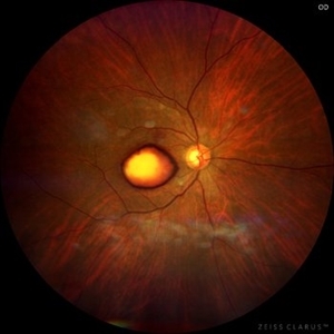 Dehemoglobinized sub-internal limiting membrane hemorrhage
Jul 29 2022 by JORGE SOBERANES
Fundus photograph of a 70-year-old man with Valsalva retinopathy manifested as premacular hemorrhage (sub-ILM) in dehemoglobinized process.
Photographer: Jorge I. Soberanes, Asociación para Evitar la Ceguera en México.
Imaging device: Zeiss Clarus 700
Condition/keywords: dehemoglobinized hemorrhage, sub-inner limiting membrane hemorrhage, valsalva retinopathy
-
 Sub ILM Dehaemoglobinised Hemorrhage With Retinal Detachment in Vitrectomised Eye
Jan 16 2025 by Anand Temkar
A 39 yrs old male was referred to us with this presentation after a month of his first vitrectomy surgery done for VH e/w. His serum homocysteine was raised but MRI brain was within normal limits. We can see the sub ILM dehaemoglobinised hemorrhage (supero-temporal to macula) and retinal detachment (inferiorly and nasally).
Photographer: Dr.Anand Temkar- Retina Foundation, Ahmedabad
Imaging device: Mirante
Condition/keywords: dehemoglobinized hemorrhage, Retinal Detachment, SUB ILM hemorrhage
-
 Sub ILM Dehaemoglobinised Hemorrhage With Retinal Detachment
Jan 16 2025 by Anand Temkar
A 39 year old male was referred to us with this presentation after a month of his first vitrectomy surgery done for VH e/w. His serum homocysteine was raised but MRI brain was within normal limits. We can see the sub ILM dehaemoglobinised hemorrhage (supero-temporal to macula) and Retinal detachment (inferiorly and nasally).
Photographer: Dr.Anand Temkar- Retina Foundation, Ahmedabad
Imaging device: Mirante
Condition/keywords: dehemoglobinized hemorrhage, Retinal Detachment, SUB ILM hemorrhage
 Loading…