Search results (466 results)
-
---thumb.jpg/image-square;max$300,300.ImageHandler) Bergmeister's Papilla
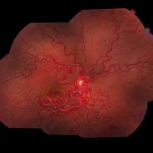
Bergmeister's Papilla
Mar 22 2014 by Hamid Ahmadieh, MD
Color fundus photograph of the right eye of a 50-year-old man with Bergmeister's papilla.
Photographer: Naghmeh Nozhat, Negah Eye Center, Tehran
Imaging device: Topcon Fundus Camera
Condition/keywords: Bergmeister's Papillae, color photo
-
 Vortex Vein Varix - supine
Dec 19 2012 by Eric A. Postel, MD
Color photograph of a vortex vein varix in a patient in the supine position
Condition/keywords: vortex vein
-
 Hollenhorst Plaque
Sep 18 2016 by John T. Thompson, MD
Color photo of Hollenhorst plaque at branch of inferotemporal artery.
Imaging device: Zeiss FF4
Condition/keywords: branch retinal artery occlusion (BRAO), hollenhorst plaque
-
 White Without Pressure
Jan 31 2018 by Olivia Rainey
Ultra-wide field pseudocolor photograph of a 57-year-old female with white without pressure affecting her left eye. Patient will be having bloodwork done to rule out possible sarcoidosis or sickle cell.
Photographer: Olivia Rainey
Imaging device: Optos
Condition/keywords: blot hemorrhages, color fundus photograph, left eye, Optos, ultra-wide field imaging, white without pressure
-
 Optic Nerve Coloboma With 2 Pits, Nasal and Temporal Color
Nov 21 2013 by Alexandre Durao Alves Pereira, MD
Fundus photograph, color, red free, blue lite and FAF of a optic nerve coloboma with 2 pits, one nasal and other temporal.
Photographer: Alexandre Pereira
Imaging device: Visucam 300
Condition/keywords: color photo, optic nerve coloboma
-
 Vortex Vein Varix - upright and invisible
Dec 19 2012 by Eric A. Postel, MD
Color photograph of a vortex vein varix in a patient in the upright position - the varix is now not visible
Condition/keywords: vortex vein
-
 Hands of 3 generations of a Marfan's family
Dec 19 2012 by Eric A. Postel, MD
Color photograph of the hands of patients from 3 generations of a family with Marfan's Syndrome.
Condition/keywords: Marfan's syndrome
-
---thumb.jpg/image-square;max$300,300.ImageHandler) Mellaril Toxicity
Mellaril Toxicity
Feb 14 2013 by From the Collections of Thomas M. Aaberg, MD and Thomas M. Aaberg Jr., MD
Composite color photography; left eye; advanced atrophy in a patient with mellaril toxicity.
Condition/keywords: mellaril toxicity
-
 Bruch’s membrane rupture
Jan 11 2013 by Hyung-Woo Kwak, MD
An area of Bruch’s membrane rupture involving the fovea is seen on color photograph (left).
Photographer: Misook Lee, Kyung Hee Univsersity Hospital, Seoul
Imaging device: Zeiss f 450 plus
Condition/keywords: myopic choroidal neovascularization (CNV)
-
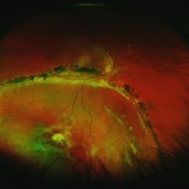 Chronic Inferior Retinal Detachment
Mar 1 2017 by Philip J. Polkinghorne, MD
Color photograph of chronic retinal detachment with pigment demarcation line and atrophic holes visible. The vision was recorded at 20/20, and follow up is 3 years.
Photographer: Alex Fraser
Condition/keywords: atrophic retinal hole, demarcation line
-
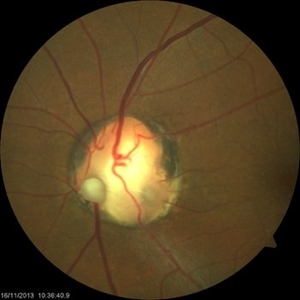
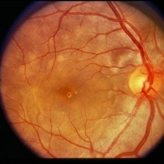
 Probable Optic Nerve Coloboma
Feb 19 2013 by From the Collections of Thomas M. Aaberg, MD and Thomas M. Aaberg Jr., MD
No history.
Condition/keywords: color photo, optic nerve coloboma
-
 Exudative Macular Degeneration, Prominent Plaque - FC
Oct 9 2012 by James B. Soque, CRA, OCT-C, COA, FOPS
88 y/o WM with extensive history of EMD and prominent plaque OS. Large heme OS has resolved with a variety of anti-VEGF therapy, and only a small heme, IT to fovea, remains. VA OS cc 20/50. (3) three photos: Color Photo, Early FA, and Late FA enclosed. 50 Degree, no mag.
Photographer: James Soque CRA COA
Imaging device: Topcon TRC 50 EX, with OIS V 10.5.74 Software. 5 MP Camera
Condition/keywords: exudative age-related macular degeneration
-
 Linear Nevus Sebaceous Syndrome
Feb 20 2015 by H. Michael Lambert, MD
Color photo of conjuctival lipodermoid in linear sebaceous nevus syndrome .
Condition/keywords: conjunctiva, linear nevus sebaceous syndrome, lipodermoid
-
 Benign Concentric Macular Annular Dystrophy
Feb 14 2013 by From the Collections of Thomas M. Aaberg, MD and Thomas M. Aaberg Jr., MD
Color photo.
Condition/keywords: benign concentric macular annular dystrophy, bull's eye maculopathy, macular dystrophy
-
---thumb.jpg/image-square;max$300,300.ImageHandler) Congenital Nyctalopia
Congenital Nyctalopia
Feb 20 2013 by From the Collections of Thomas M. Aaberg, MD and Thomas M. Aaberg Jr., MD
Color photo of the periphery showing RPE mottling, clumping, and small multiple yellow deposits.
Condition/keywords: color photo, congenital nyctalopia, retinal pigment epithelium
-
 Retinoschisis Right Eye
Oct 27 2014 by AnneMarie Smykowski
65-year-old white male, with hypertensive retinopathy. He has a stable retinoschisis for approximately 10 years.
Photographer: AnneMarie Smykowski C.O.A., Island Retina Shirley, NY
Imaging device: Optos Daytona
Condition/keywords: color photo, Daytona, Optos, retinoschisis
-
---thumb.jpg/image-square;max$300,300.ImageHandler) Flexibility in Marfan's Syndrome
Flexibility in Marfan's Syndrome
Dec 19 2012 by Eric A. Postel, MD
Color photo demonstrating flexibility and long fingers in a patient with Marfan's Syndrome
Condition/keywords: Marfan's syndrome
-
 Lattice Degeneration With Atrophic Hole
Feb 19 2015 by H. Michael Lambert, MD
Color photo of Lattice degeneration with atrophic hole.
Condition/keywords: atrophic retinal hole, lattice degeneration
-
 Exudative Macular Degeneration, Prominent Plaque - FA early
Oct 9 2012 by James B. Soque, CRA, OCT-C, COA, FOPS
88 y/o WM with extensive history of EMD and prominent plaque OS. Large heme OS has resolved with a variety of anti-VEGF therapy, and only a small heme, IT to fovea, remains. VA OS cc 20/50. (3) three photos: Color Photo, Early FA, and Late FA enclosed. 50 Degree, no mag.
Photographer: James Soque CRA COA
Imaging device: Topcon TRC 50 EX, with OIS V 10.5.74 Software. 5 MP Camera
Condition/keywords: exudative age-related macular degeneration
-
---thumb.jpg/image-square;max$300,300.ImageHandler) Optic And Hyperemic Disc And Hyperemic Rim
Optic And Hyperemic Disc And Hyperemic Rim
Nov 4 2013 by Maurice F. Rabb
Color photographs of the right posterior pole demonstrates an optic disc with a cup/disc ratio of 0.5 and a hyperemic disc and hyperemic rim of neutral tissue.
Condition/keywords: hyperemic disc, hyperemic rim, optic disc
-
 Myelinated Nerve Fibers and Possibly Posterior Staphyloma
Feb 19 2013 by From the Collections of Thomas M. Aaberg, MD and Thomas M. Aaberg Jr., MD
Findings are bilateral.
Condition/keywords: color photo, myelinated nerve fibers
-
---thumb.jpg/image-square;max$300,300.ImageHandler) Cone Dystrophy
Cone Dystrophy
Feb 20 2013 by From the Collections of Thomas M. Aaberg, MD and Thomas M. Aaberg Jr., MD
Color photo of the fundus of OD in a patient with cone dystrophy; VA=20/80.
Condition/keywords: color photo, cone dystrophy, macula
-
 Wyburn Mason Racemose Angiomatosis
May 22 2016 by Olivia Rainey
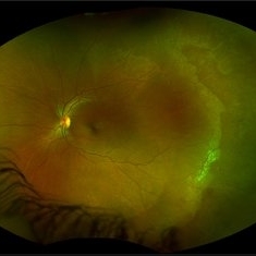
Color fundus montage of an 13-year-old female with arteriovenous malformation (Wyburn Mason Racemose Angiomatosis) affecting her right eye. The retinal arteriovenous malformation appears to be stable. She presented with NLP in the eye, strabismus, and peripheral retinal ischemia. She is at risk for neovascular complications; however, she is currently being treated with Sirolimus. Since she is on this systemically, there is no need to perform intraocular anti-VEGF injections or PRP laser. She also presented with optic atrophy affecting her left eye, secondary to chiasmal involvement of arteriovenous malformation. She has had a potential progressive visual field loss involving the temporal aspect of her visual field from the left eye. There is sector optic atrophy. Presumably, this is due to a compressive effect of her arteriovenous malformation on the nasal nerve fiber layer (corresponding to the temporal visual field) crossing to the right occipital cortex at the chiasm.
Photographer: Olivia Rainey
Imaging device: Topcon 50dx
Condition/keywords: arteriovenous malformation, color fundus photograph, color photo, montage, peripheral ischemia, Sirolimus
-
 Retinoschisis Left Eye
Oct 27 2014 by AnneMarie Smykowski
65-year-old white male, with hypertensive retinopathy. He has a stable retinoshisis for approximately 10 years.
Photographer: AnneMarie Smykowski C.O.A., Island Retina Shirley, NY
Imaging device: Optos Daytona
Condition/keywords: color photo, Daytona, Optos, retinoschisis
-
 Vogt-Koyanagi-Harada Disease
Feb 20 2015 by H. Michael Lambert, MD
Color photo showing multifocal detachments of the neurosensory retina with underlying cream colored lesions ( possibly Dalen-Fuchs nodules). Large pocket of subretinal fluid in the macula.
Condition/keywords: exudative retinal detachment, Vogt-Koyanagi-Harada
 Loading…