Search results (24 results)
-
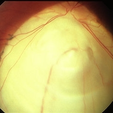
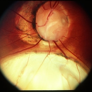 Coloboma
Mar 29 2013 by Henry J. Kaplan, MD
Optic disc and inferonasal choroidal coloboma in the same patient #2.
Condition/keywords: coloboma, coloboma of choroid, coloboma of optic disc
-
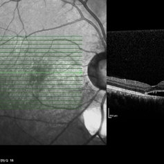
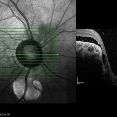 Coloboma of Disc & Choroid
Oct 6 2012 by Hamid Ahmadieh, MD
OCT image of a 25-year-old woman with serous retinal detachment secondary to coloboma of disc associated with coloboma of choroid.
Photographer: Hamid Ahmadieh, MD, Ophthalmic Research Center, Labbafinejad Medical Center, Shahid Beheshti University of Medical Sciences
Imaging device: Heidelberg Spectralis
Condition/keywords: coloboma of choroid, coloboma of optic disc, optical coherence tomography (OCT), serous retinal detachment
-
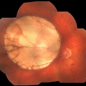
 Coloboma
Mar 29 2013 by Henry J. Kaplan, MD
Large choroidal coloboma.
Condition/keywords: coloboma, coloboma of choroid
-
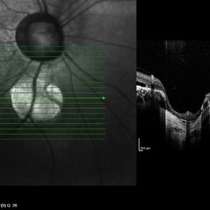 Coloboma of Disc & Choroid
Oct 6 2012 by Hamid Ahmadieh, MD
OCT image of a 25-year-old woman with a small coloboma of choroid associated with coloboma of disc.
Photographer: Hamid Ahmadieh, MD, Ophthalmic Research Center, Labbafinejad Medical Center, Shahid Beheshti University of Medical Sciences
Imaging device: Heidelberg Spectralis
Condition/keywords: coloboma of choroid, coloboma of optic disc, optical coherence tomography (OCT)
-
 Coloboma of Disc & Choroid
Oct 6 2012 by Hamid Ahmadieh, MD
OCT image of a 25-year-old woman with serous retinal detachment secondary to coloboma of disc associated with coloboma of choroid.
Photographer: Hamid Ahmadieh, MD, Ophthalmic Research Center, Labbafinejad Medical Center, Shahid Beheshti University of Medical Sciences
Imaging device: Heidelberg Spectralis
Condition/keywords: coloboma of choroid, coloboma of optic disc, optical coherence tomography (OCT), serous retinal detachment
-
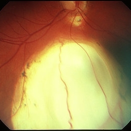
 Chorioretinal coloboma 1
Jan 11 2013 by Alex P. Hunyor, MD
Inferior chorioretinal coloboma - color image 1.
Condition/keywords: chorioretinal coloboma, coloboma of choroid
-
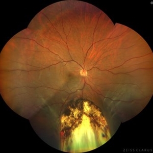
 Coloboma
Mar 29 2013 by Henry J. Kaplan, MD
Coloboma involving optic nerve and inferior choroid.
Condition/keywords: coloboma of choroid, coloboma of optic disc
-
 Chorioretinal coloboma 2
Jan 11 2013 by Alex P. Hunyor, MD
Inferior chorioretinal coloboma - color image 2.
Condition/keywords: chorioretinal coloboma, coloboma of choroid
-
 Coloboma
Jan 23 2018 by JEFFERSON R SOUSA, Tecg.º (Biomedical Systems Technology)
Male patient, 22 years old, with low vision since infancy. In retinal and retinal mapping examinations, important alterations were observed in the formation of retinochoroidal structures suggestive of coloboma.
Photographer: JEFFERSON R SOUSA - Study Center and Ophthalmological Research Dr. Andre M V Gomes, Dr. Suel Abujamra Institute São Paulo-Brazil
Imaging device: Acquisition of the image in the Camera background Topcon TRC-50 Dx - IA, Keystone field photo of 50 Degrees. Composition automatic of Imaginet with manual adjustment
Condition/keywords: coloboma, coloboma of choroid
-
 Coloboma
Oct 2 2019 by John S. King, MD
27-year-old white female with bilateral, isolated, inferior, chorioretinal colobomas; she has a history of retinal laser anterior to the edge of the coloboma OD secondary to a limited RD. This is the right eye.
Photographer: Shelly Blair
Imaging device: Optos CA
Condition/keywords: coloboma of choroid
-
 Dislocated IOL in Vitreous in a Eye With Choroidal Coloboma
Jan 24 2018 by Manish Nagpal, MD, FRCS (UK), FASRS
60-year-old male patient had come with sudden decrease in vision post trauma with hand and on examination the IOL had dislocated in the vitreous alongside a choroidal coloboma.
Photographer: Mehul Prajapati
Condition/keywords: coloboma of choroid, dislocated posterior chamber intraocular lens (PCIOL)
-
 Coloboma
Sep 7 2018 by John S. King, MD
Bilateral retinochoroidal colobomas in 29-year-old white female with 20/50 OD and 20/40 OS acuity and bilateral inferior iris colobomas. No history of RD.
Photographer: KO
Imaging device: Optos CA
Condition/keywords: coloboma of choroid
-
 Coloboma
Oct 2 2019 by John S. King, MD
27-year-old white female with bilateral, isolated, inferior, chorioretinal colobomas; she has a history of retinal laser anterior to the edge of the coloboma OD secondary to a limited RD. This is the left eye.
Photographer: Shelly Blair
Imaging device: Optos CA
Condition/keywords: coloboma of choroid
-
 Coloboma involving the Optic nerve, Retina, and Choroid
Dec 6 2021 by Jesus Lozano, MD
78-year-old woman after prophylactic laser photocoagulation (PLP) for her RE Coloboma involving the optic nerve, retina, and choroid. At 6 month follow up, patient preserved her FC vision as it was before the procedure. Retina attached.
Photographer: Yair Bet Yosef, Hadassah Medical Center. Israel
Imaging device: Optos Silverstone fundus image
Condition/keywords: coloboma, coloboma of choroid, coloboma of macula, coloboma of optic disc, PLP, prophylactic photocoagulation
-
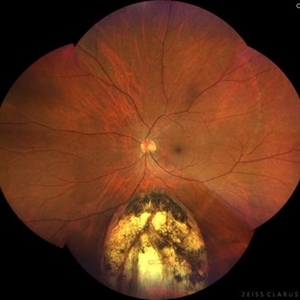
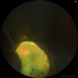
 Retinal detachment, Coloboma
Oct 17 2022 by JEFFERSON R SOUSA, Tecg.º (Biomedical Systems Technology)
Male patient, 58 years old, PIO 19-21, AV; OD 20/200, OS 20/20. He had sudden low vision, with progressive worsening in his right eye. He underwent evaluation in retinal mapping and further examinations where important funduscopic changes were found. Both eyes had an atrophic lesion suggestive of coloboma of the retina in the lower arch. Right eye with retinal detachment with macular involvement.
Photographer: JEFFERSON ROCHA DE SOUSA - Retinal Department at Instituto Dr. Suel Abujamra Sao Paulo-Brazil
Imaging device: Clarus 700 - Zeiss, composite of four 135 degree images.
Condition/keywords: coloboma, coloboma of choroid, Retinal Detachment
-
 Optic Disc Pit Associated with Optic Disc Coloboma and Retinochoroidal Coloboma
Jul 22 2020 by Deepak Bhojwani, MS
Fundus photograph of a 32-year-old male showing large optic disc pit in a colobomatous optic nerve head along with isolated inferior retino-choroidal coloboma. (A rare / coincidental occurrence of multiple congenital anomalies of optic disc and retina)
Photographer: DEEPAK BHOJWANI
Condition/keywords: coloboma of choroid, coloboma of optic disc, congenital optic nerve pit
-
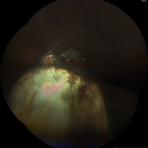
 Retinal detachment
Oct 17 2022 by JEFFERSON R SOUSA, Tecg.º (Biomedical Systems Technology)
Male patient, 58 years old, PIO; 19-21, AV; OD 20/200, OS 20/20. He had sudden low vision, with progressive worsening in his right eye. He underwent evaluation in retinal mapping and further examinations where important funduscopic changes were found. Both eyes had an atrophic lesion suggestive of coloboma of the retina in the lower arch. Right eye with retinal detachment with macular involvement.
Photographer: JEFFERSON ROCHA DE SOUSA - Retinal Department at Instituto Dr. Suel Abujamra Sao Paulo-Brazil
Imaging device: Clarus 700 - Zeiss, composite of four 135 degree images.
Condition/keywords: coloboma, coloboma of choroid, Retinal Detachment
-
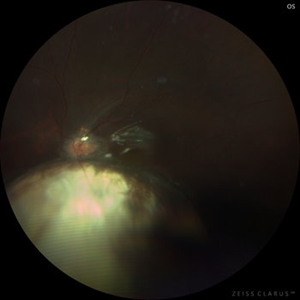
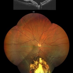 Retinal detachment, Coloboma
Oct 17 2022 by JEFFERSON R SOUSA, Tecg.º (Biomedical Systems Technology)
Male patient, 58 years old, PIO; 19-21, AV; OD 20/200, OS 20/20. He had sudden low vision, with progressive worsening in his right eye. He underwent evaluation in retinal mapping and further examinations where important funduscopic changes were found. Both eyes had an atrophic lesion suggestive of coloboma of the retina in the lower arch. Right eye with retinal detachment with macular involvement.
Photographer: JEFFERSON ROCHA DE SOUSA - Retinal Department at Instituto Dr. Suel Abujamra Sao Paulo-Brazil
Imaging device: Clarus 700 - Zeiss, composite of four 135 degree images. CIRRUS 5000, Protocol, HD 5 Line. con
Condition/keywords: coloboma of choroid, Retinal Detachment
-
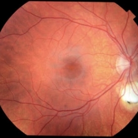 Bilateral Coloboma-OD
Apr 29 2022 by Deepika Malik, MD
A adolescent male with a history of intellectual disability was referred for ophthalmologic evaluation of monocular vision loss. Fundoscopic examination of the right eye revealed an inferior typical retinochoroidal coloboma without involvement of the disc. The left eye revealed an optic pit within an inferior retinochoroidal coloboma leading to a localized retinal detachment.
Condition/keywords: coloboma of choroid
-
 Restoring the glow : Retinochoroidal Coloboma associated Retinal detachment - Pre-op
Aug 21 2023 by Harsh Vardhan Singh, MS
17 year-old female with RC coloboma with total bullous retinal detachment
Photographer: Dr Harsh Vardhan Singh
Condition/keywords: coloboma of choroid, pre-op
-
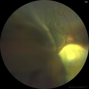
 Disc Coloboma
Aug 17 2023 by Dr.Anushri Godbole
21 years old female came to OPD with chief complaints of diminution of vision of LE since birth. BCVA RE-6/6 N6, LE FC-1/2M, N36. On examination RE was diagnosed as disc coloboma with type 6 coloboma in periphery and LE was diagnosed as Choroidal coloboma involving disc
Condition/keywords: coloboma of choroid, coloboma of optic disc
-
 Restoring the glow : Retinochoroidal Coloboma associated Retinal detachment - Pre-op
Aug 21 2023 by Harsh Vardhan Singh, MS
17-year-old female with RC coloboma with total bullous retinal detachment
Photographer: Dr Harsh Vardhan Singh
Condition/keywords: coloboma of choroid, pre-op
-
 Restoring the glow: Retinochoroidal Coloboma associated Retinal detachment - Post-op
Aug 21 2023 by Harsh Vardhan Singh, MS
17 year-old female with RC coloboma with total bullous retinal detachment - operated for the same
Photographer: Dr Harsh Vardhan Singh
Condition/keywords: coloboma of choroid, post-op
 Loading…